KOSIN UNIVERSITY COLLEGE OF MEDICINE
Articles
- Page Path
- HOME > Kosin Med J > Volume 35(1); 2020 > Article
-
Case Reports
Atypical Lipomatous Tumor/Well-Differentiated Liposarcoma Arising from the Tongue: case report - Young Chul Kim, Somi Ryu, Seong Jun Won, Jung Je Park
-
Kosin Medical Journal 2020;35(1):58-63.
DOI: https://doi.org/10.7180/kmj.2020.35.1.58
Published online: June 30, 2020
1Department of Otolaryngology, Gyeongsang National University School of Medicine, Gyeongsang National University Hospital, Jinju, Korea
2Institute of Health Sciences, College of Medicine, Gyeongsang National University, Jinju, Korea
- Corresponding Author: Jung Je Park, Department of Otorhinolaryngology, Head & Neck Surgery and Institute of Health Sciences, Gyeongsang National University School of Medicine, Gyeongsang National University Hospital, 501, Jinju-daero, Jinju 52828, Korea, Tel: +82-55-750-8698, Fax: +82-55-759-0613, E-mail: capetown@hanmail.net
• Received: December 23, 2019 • Revised: April 17, 2020 • Accepted: May 6, 2020
Copyright © 2020 by Korean Association of Medical Journal Editors
Articles published in Kosin Medical Journal are open-access, distributed under the terms of the Creative Commons Attribution Non-Commercial License (https://creativecommons.org/licenses/by-nc/4.0/) which permits unrestricted non-commercial use, distribution, and reproduction in any medium, provided the original work is properly cited.
- 1,204 Views
- 7 Download
Abstract
- Liposarcomas are common mesenchymal malignant tumors arising from adipose tissue. Although liposarcomas are the most frequent type of soft tissue sarcomas, accounting for approximately 20% of all soft tissue sarcomas, they are rare in the head and neck, particularly in the oral cavity. Oral liposarcomas have been reported to occur mainly on the buccal mucosa, with other sites including the floor of the mouth, tongue, palate, and mandible. This report describes a 76-year-old male patient with an atypical lipomatous tumor/well-differentiated liposarcoma of the tongue that underwent surgical excision. This report also reviews published data on these rare tumors.
- A 76-year-old male patient presented with an asymptomatic, slowly growing tongue mass of unclear onset. The patient had undergone treatment for hypertension, diabetes, and chronic renal failure and was a 30 pack-year ex-smoker. Physical examination revealed a firm cystic mass with a smooth surface, approximately 1 × 1 cm in size, in the right anterior aspect of the tongue (Fig. 1). There was no evidence of cervical lymphadenopathy. Preoperative T1-weighted magnetic resonance imaging (MRI) of the tongue showed a well-defined, ovoid-shaped mass with high-signal intensity, with T1-enhanced MRI showing attenuated signal intensity inside the lesion (Fig. 2). Cervical computed tomography (CT) revealed no cervical lymph node enlargement or other specific findings.
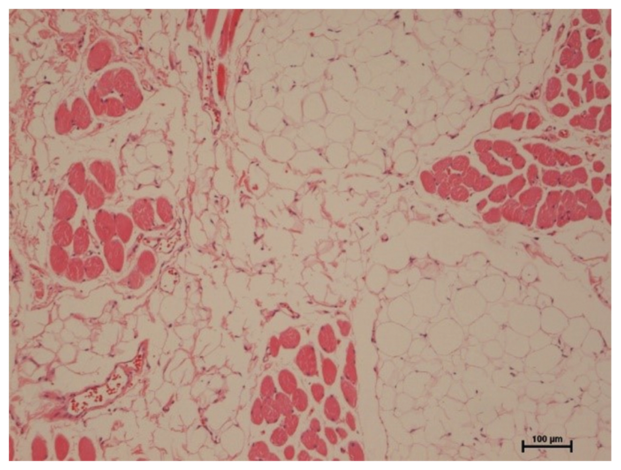
- The patient was provisionally diagnosed with a lipoma of the tongue, which was removed surgically. Histopathological examination of the lesion revealed a well-differentiated liposarcoma, also called an atypical lipomatous tumor, with undetermined resection margins. Histologically, the tumor was mainly composed of fatty cells with variation in size and shape and intervening fibrous septum. It also shows characteristic lipoblasts with enlarged indented nuclei and variously sized lipid-containing cytoplasmic vacuoles (Fig. 3).2
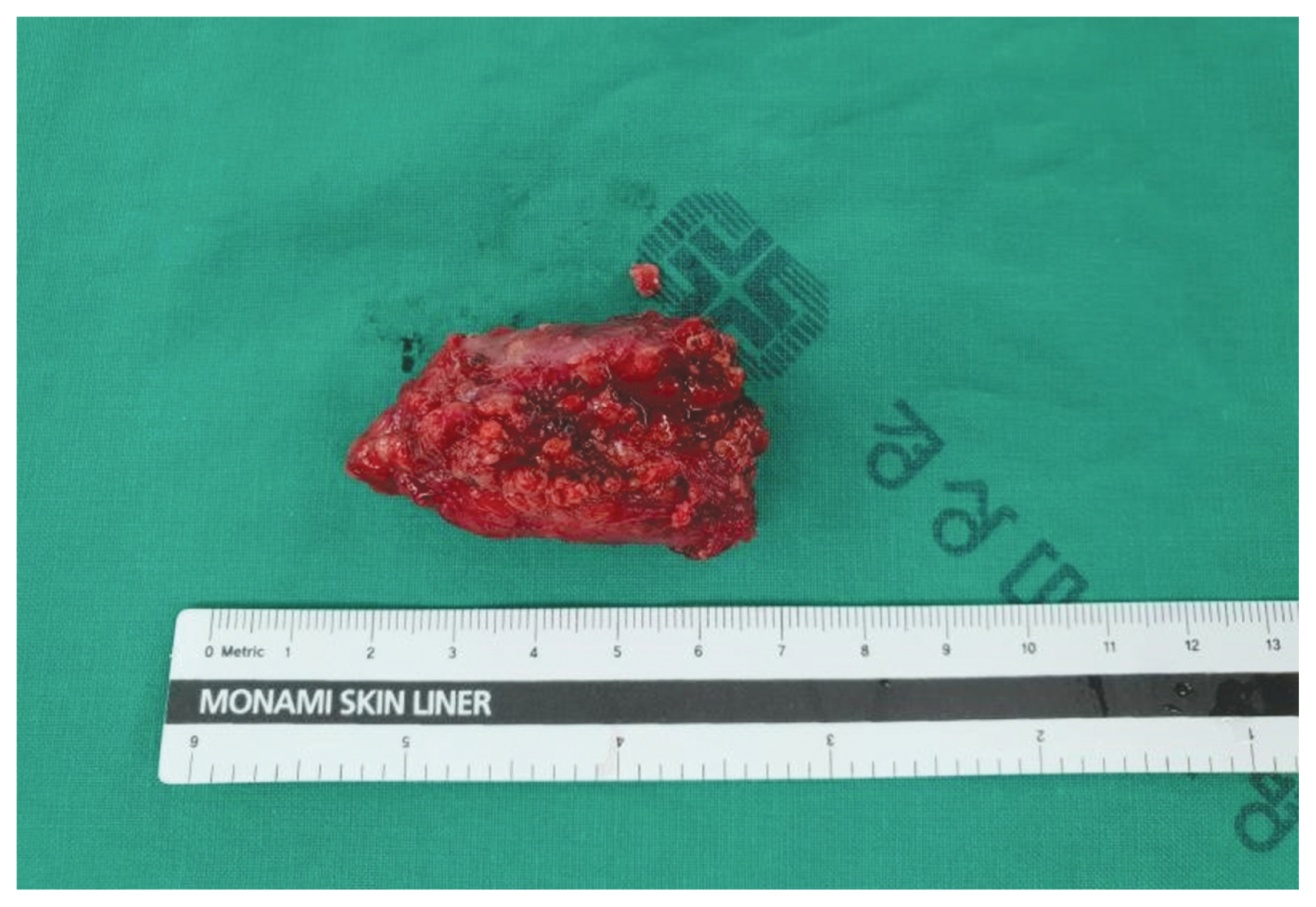
- (Fig. 3). The patient was assessed for metastases by positron emission tomography (PET)-CT, abdominal sonography, and gastroscopy, but no evidence of metastasis was observed. A second operation was performed to completely resect the tumor, including safety margins. The postoperative course of the patient was uneventful, with no evidence of tumor recurrence 1 year later.
CASE
- Liposarcomas, first described in 1857, account for approximately 20% of all soft tissue malignancies and are the most common type of sarcoma in adults. Liposarcomas may arise wherever adipose tissue is normally present, but most occur in deep soft tissues and the retroperitoneum. 3,4 Head and neck liposarcomas are uncommon, constituting 1.8 – 6% of all liposarcomas. Oral liposarcomas are extremely rare. The most common sites in the oral cavity for their occurrence are the oral mucosa (38%), the tongue (33%). the floor of the mouth (7%) and the palate (7%).4
- Liposarcomas have been classified into several types, including well-differentiated, dedifferentiated, myxoid, pleomorphic and round cell types. Myxoid type is the most common (30 – 50%), followed by well differentiated (20 – 30%), pleomorphic (10 – 25%) and round cell (10 – 15%) types.5 Well-differentiated liposarcomas, especially in the oral cavity, are predominantly superficial, are of small size, and have clear margins. These tumors rarely metastasize or recur. The WHO has classified well-differentiated liposarcomas as atypical lipomatous tumors. 1 The 5-year survival rate of patients with atypical lipomatous tumors was reported to range from 75% to 100%, compared with a 20% 5-year survival rate in patients with pleomorphic and round cell type tumors.6
- The distinction between lipoma and atypical lipomatous tumor, however, is a frequent diagnostic dilemma.2 Although these tumors are firmer and less easily compressed and fixed than benign lipomas, they are not easy to distinguish clinically.7 The diagnosis of atypical lipomatous tumors is based on pathologic findings, including the presence of lipoblasts, variations in adipocyte size, and adipocytes with atypical and enlarged nuclei.8,9 Liposarcoma of the tongue can easily be mistaken for benign lipoma, traumatic fibroma, papilloma, generalized gingiva hyperplasia, and pyogenic granuloma.2
- Surgical resection is the treatment of choice.7 Wide excision, including safety margins, can significantly reduce recurrence rates.9 However some groups recommended conservative surgical excision with close and long periods of follow–up instead of radical surgical approach due to a generally favorable prognosis based on the histopathologic subtype, location and clear surgical margins.10 Local recurrence rates are reported to range from 7% to 30% due to incomplete resection.4,11 Postoperative radiation therapy may prevent tumor recurrence, especially of radiation sensitive myxoid type tumors. Postoperative radiation therapy is not required for atypical lipomatous tumors if resection margins are tumor free and there is no capsular invasion.12,13 The patient described in this report did not receive additional radiation therapy because there was no involvement of the capsule after wide excision. The average size of liposarcomas of the tongue is 0.8 cm, with prognosis being better in patients with tumors < 5 cm in size.14 By contrast, patients with liposarcomas arising in the retroperitoneum or trunk have a poor prognosis due to the difficulty of surgical resection.4,15
- Despite its unclear onset, the tumor in this patient was thought to be benign because of its size and characteristics, including its cystic appearance and smooth surface on palpation. Furthermore, imaging suggested that it was a lipoma. However, histologic examination of the lesion suggested that it was an atypical lipomatous tumor. The diagnosis is made histologically. 12 These tumors should be actively treated if their duration is long or unclear and/or they are large in size. In summary, this report described a patient with a rare atypical lipomatous tumor/well-differentiated liposarcoma of the tongue. The tumor was successfully surgically removed, with no evidence of recurrence at 1-year follow up.
DISCUSSION
Fig. 1Laryngoscopic finding, showing a 1 × 1cm sized yellowish mass arising from the right anterior lateral tongue. The mass had smooth surfaces and was well encapsulated.
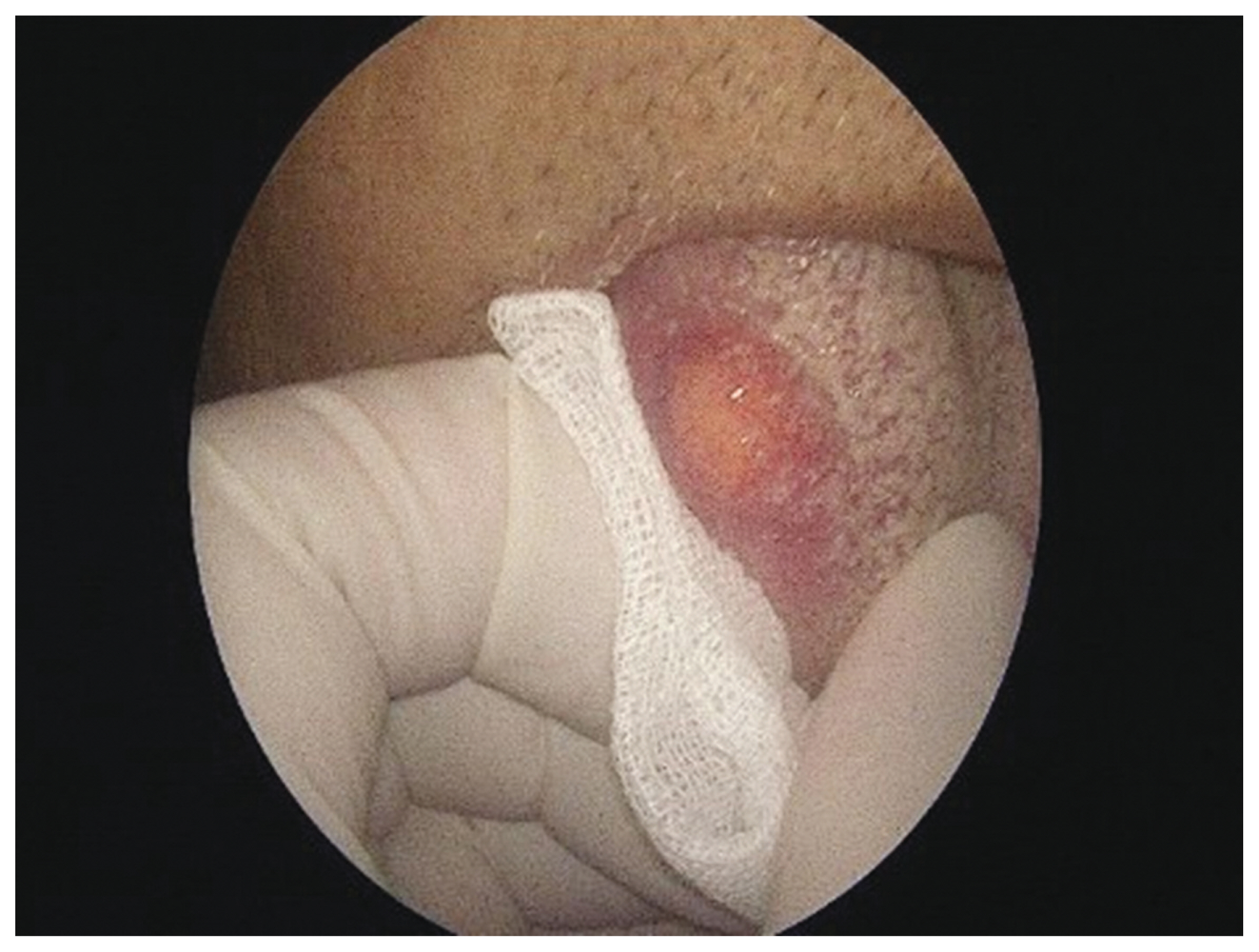
Fig. 2Preoperative magnetic resonance imaging (MRI) of the tongue mass. (A) Transverse T1-weighted MRI showing a well-defined, ovoid-shaped mass with homogeneous high-signal intensity. (B) Transverse, fat-suppressed T1-weighted, gadolinium-enhanced MRI showing attenuated signal intensity inside the lesion.
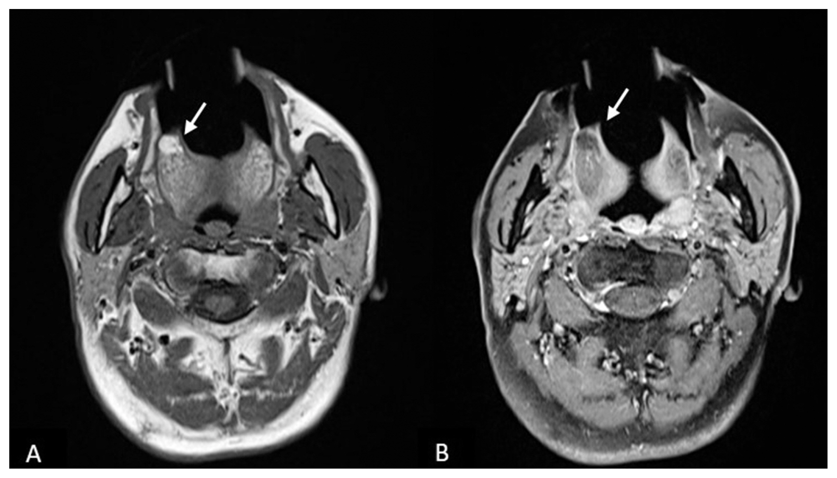
Fig. 3Pathologic finding. An atypical lipomatous tumor showing mature adipocytes with great variations in size and shape in fibrous stroma (H&E, ×100)

Fig. 4Lesion removed during the second operation. The specimen was about 6 x 2.5 x 1 cm in size, including a 2 cm safety margin.
- 1. Nascimento AF, McMenamin ME, Fletcher CD. Liposarcomas/atypical lipomatous tumors of the oral cavity: a clinicopathologic study of 23 cases. Ann Diagn Pathol 2002;6:83–93.ArticlePubMed
- 2. Kim YB, Leem DH, Baek JA, Ko SO. Atypical lipomatous tumor/well-differentiated liposarcoma of the gingiva: a case report and review of literature. J Oral Maxillofac Surg 2014;72:431–9.ArticlePubMed
- 3. Laco J, Mentzel T, Hornychova H, Kohout A, Jirousek Z, Ryska A. Atypical lipomatous tumors of the tongue: report of six cases. Virchows Arch 2009;455:383–8.ArticlePubMed
- 4. DeWitt J, Heidelman J, Summerlin DJ, Tomich C. Atypical lipomatous tumors of the oral cavity: a report of 2 cases. J Oral Maxillofac Surg 2008;66:366–9.ArticlePubMed
- 5. Yeung MJ, Serpell JW. Management of the solitary thyroid nodule. Oncologist 2008;13:105–12.ArticlePubMed
- 6. Davis EC, Ballo MT, Luna MA, Patel SR, Roberts DB, Nong X, et al. Liposarcoma of the head and neck: The University of Texas M. D. Anderson Cancer Center experience. Head Neck 2009;31:28–36.ArticlePubMed
- 7. Demir D, Katircioglu S, Suoglu Y, Bilgic B. Radiation-induced liposarcoma of the retropharyngeal space. Otolaryngol Head Neck Surg 2006;134:1060–2.ArticlePubMed
- 8. Nunes FD, Loducca SV, de Oliveira EM, de Araujo VC. Well-differentiated liposarcoma of the tongue. Oral Oncol 2002;38:117–9.ArticlePubMed
- 9. Casani AP, Marchetti M, Dallan I, Cagno MC, Berrettini S. Liposarcoma of the cervico-nuchal region. Otolaryngol Head Neck Surg 2005;133:641.Article
- 10. Nili F, Baghai F, Aghai A, Etebarian A. Well-differentiated liposarcoma of the floor of the mouth: report of a rare case and review of the literature. J Oral Maxillofac Pathol 2016;20:312–5.ArticlePubMedPMC
- 11. Piperi E, Tosios KI, Nikitakis NG, Kyriakopoulos VF, Tzerbos F, Koutlas IG, et al. Well-differentiated liposarcoma/atypical lipomatous tumor of the oral cavity: report of three cases and review of the literature. Head Neck Pathol 2012;6:354–63.ArticlePubMedPMC
- 12. Miyazaki M, Aoki M, Oba S, Sakata T, Nakagawa T, Nabeshima K. A rare case of dedifferentiated liposarcoma of the sinonasal cavity: a case report. Mol Clin Oncol 2017;7:539–42.ArticlePubMedPMC
- 13. Favia G, Maiorano E, Orsini G, Piattelli A. Myxoid liposarcoma of the oral cavity with involvement of the periodontal tissues. J Clin Periodontol 2001;28:109–12.ArticlePubMed
- 14. Fanburg-Smith JC, Furlong MA, Childers EL. Liposarcoma of the oral and salivary gland region: a clinicopathologic study of 18 cases with emphasis on specific sites, morphologic subtypes, and clinical outcome. Mod Pathol 2002;15:1020–31.ArticlePubMed
- 15. Crago AM, Singer S. Clinical and molecular approaches to well differentiated and dedifferentiated liposarcoma. Curr Opin Oncol 2011;23:373–8.ArticlePubMedPMC
References
Figure & Data
References
Citations
Citations to this article as recorded by 
 PubReader
 ePub Link
 Cite
Cite