KOSIN UNIVERSITY COLLEGE OF MEDICINE
Articles
- Page Path
- HOME > Kosin Med J > Volume 38(3); 2023 > Article
-
Original article
Post-percutaneous core needle biopsy sputum cytology: diagnostic value and factors for positive prediction in diagnosing malignancy -
Sang Kyu Lee1
 , Hee Kang1, Min Jung Jung2, Sekyoung Park1, Ki Nam Lee1
 -
Kosin Medical Journal 2023;38(3):201-209.
DOI: https://doi.org/10.7180/kmj.23.127
Published online: August 16, 2023
1Department of Radiology, Kosin University Gospel Hospital, Kosin University College of Medicine, Busan, Korea
2Department of Pathology, Kosin University Gospel Hospital, Kosin University College of Medicine, Busan, Korea
- Corresponding Author: Hee Kang, MD, PhD Department of Radiology, Kosin University Gospel Hospital, Kosin University College of Medicine, 262 Gamcheon-ro, Seo-gu, Busan 49267, Korea Tel: +82-51-990-6341 Fax: +82-51-255-2764 E-mail: kanghi81@gmail.com
Copyright © 2023 Kosin University College of Medicine.
This is an open-access article distributed under the terms of the Creative Commons Attribution Non-Commercial License (http://creativecommons.org/licenses/by-nc/4.0/) which permits unrestricted non-commercial use, distribution, and reproduction in any medium, provided the original work is properly cited.
- 1,184 Views
- 13 Download
Abstract
-
Background
- This study evaluated the diagnostic yield and positive predictive factors of post-percutaneous core needle biopsy (PCNB) sputum cytology in diagnosing malignancy.
-
Methods
- This retrospective study included patients who underwent PCNB at a single center from January 2014 to March 2022. Patient demographics, lung lesion characteristics on computed tomography, underlying lung disease, post-PCNB complications, histopathologic results of PCNB, post-PCNB sputum specimens, and final diagnoses were reviewed. The diagnostic yields and related factors were analyzed.
-
Results
- Overall, 177 consecutive patients with sputum specimens obtained after PCNB for intrapulmonary lesions were enrolled. Among them, 152 patients had a final diagnosis of malignancy. Diagnostic sputum specimens with atypical or malignant cells were obtained in 12 patients. The sensitivity, specificity, accuracy, positive predictive value, and negative predictive value of sputum cytology were 7.89%, 100%, 20.90%, 100%, and 15.15%, respectively. Lesion size, air-bronchogram, lesion multiplicity, and the cell type of squamous cell and adenocarcinoma differed significantly between the groups with diagnostic versus non-diagnostic sputum (p<0.05). The lesion size (odds ratio [OR], 1.035; 95% confidence interval [CI], 1.008–1.064; p=0.013), presence of air-bronchogram (OR, 23.485; 95% CI, 2.532–217.316; p=0.005), and squamous cell carcinoma (OR, 7.397; 95% CI, 1.773–30.865; p=0.006) were significantly associated with a diagnostic sputum specimen post-PCNB.
-
Conclusions
- Although post-PCNB sputum cytology had low sensitivity in diagnosing lung cancer, it showed diagnostic results in some peripheral lung cancer patients who have squamous cell types, relatively large tumors, and air-bronchograms in the lesions.
- Worldwide, lung cancer is the second most common cancer and the leading cause of cancer mortality. In 2020, 2.21 million people were newly diagnosed with lung cancer, and 1.8 million died from lung cancer [1]. The crude incidence rate of lung cancer in Korea in 2019 was 58.4 per 100,000 people, making it the second highest of all cancers, and the 5-year relative survival rate of lung cancer patients was only 34.7%. Characteristically, lung cancer had the highest proportion of patients diagnosed at a distant metastatic stage, approximately 50%, with a corresponding 5-year survival rate of 7.6% [2].
- To reduce the mortality rate of lung cancer, early diagnosis is essential. In the 1970s and 1980s, large-scale trials using chest X-rays and sputum cytology for lung cancer screening failed to significantly reduce lung cancer mortality [3-5]. Since the 1990s, efforts have been made to diagnose lung cancer early using low-dose chest computed tomography (LDCT). In 2011, the National Lung Screening Trial reported a 20% reduction in lung cancer mortality from screening with LDCT [6]. However, using LDCT for lung cancer screening has low specificity, i.e., its ability to differentiate between benign and malignant lesions is poor [6,7]. Invasive pathological confirmation is imperative to diagnose malignancy in many cases.
- Sputum cytology is an examination widely used for the respiratory system because of its cost-effectiveness and non-invasiveness. The detection of atypical cells in sputum can also be helpful in diagnosing cancer. Furthermore, biomolecular analyses of sputum cytology show potential for risk assessment and the early detection of lung cancer [8]. However, sputum cytology plays a limited role because of its low sensitivity [9]. The many factors that influence the sensitivity of sputum cytology include the location, stage, and cytologic subtype of the tumor; the number of sputum specimens; the specimen-collection method; and the specimen preparation technique [10].
- Percutaneous core needle biopsy (PCNB) is a minimally invasive procedure for the pathological confirmation of peripheral lung lesions; it has high sensitivity and specificity of around 90% [11]. PCNB is a technique widely used for diagnosing lung cancer, although its value is limited in some cases of central lung cancer.
- Our purpose in this study was to evaluate the diagnostic value of sputum cytology after PCNB for lung cancer diagnosis and to investigate which factors influence the results.
Introduction
- Ethical statements: This study was approved by the Institutional Review Board (IRB) of Kosin University Gospel Hospital (IRB No: KUGH 2023-02-004). The requirement for patient consent was waived due to the retrospective nature of this study.
- 1. Study population
- Under approval of institutional review board, we retrospectively reviewed the medical records of 987 consecutive patients who underwent PCNB of the thorax in a single center from January 2014 to March 2022. Patients who underwent PCNB for extrapulmonary lesions, including pleura and chest wall lesions (n=19); those from whom no sputum could be obtained after PCNB (n=778); and those without a pathologically or clinically confirmed diagnosis (n=13) were excluded from enrollment. Therefore, 177 consecutive patients from whom sputum specimens were obtained after PCNB for the diagnosis of intrapulmonary lesions were included. We reviewed their medical records to collect demographic information (age, sex, and history of smoking), the results of pulmonary function testing, operator’s procedure record, results of post-PCNB sputum specimens, results of PCNB, and final diagnoses.
- 2. CT acquisition and assessment
- Computed tomography (CT) scans were acquired using a multidetector CT system (Somatom Sensation 64 or dual-source Somatom Definition Flash 128 or dual-source Somatom Force 192 multidetector CT system; Siemens Medical Solutions) with or without intravenous administration of contrast medium. Scanning parameters were 80–120 kVp, 90–150 mA, 0.5 seconds tube rotation time, and 1.2 pitch. The image data were reformatted with 1- to 2-mm slice thickness for transverse images and a 2.0-mm slice thickness for coronal and sagittal images. Two radiologists with 2 and 15 years of experience in chest CT interpretation retrospectively reviewed preprocedural CT images by consensus. Images were displayed with a lung window setting of level –500 Hounsfield units (HU) and width 200 HU and mediastinal window of level 25 HU and width 40 HU. One-dimensional size measurements were performed on the maximum diameter of the lesion on axial images. We divided the location of lung lesions into central and peripheral lesions. Central lung lesions were limited to the trachea, bronchi, or segmental bronchi, and peripheral lesions were found farther to the periphery than the subsegmental bronchi. The distance from the pleura to the lesion on the CT scan was also measured. We evaluated lesion solidity according to the Fleischner 2017 guideline. Solid nodules had homogenous soft-tissue attenuation, part-solid nodules had both ground-glass and solid soft-tissue attenuation components, and pure ground glass nodules had only ground glass attenuation [12]. Other characteristics (the presence or absence of necrosis, cavity, air-bronchogram, mediastinal lymphadenopathy, multiplicity of lung lesions, and underlying lung disease of emphysema or fibrosis) were also reviewed. Necrosis was defined as low-attenuated, non-enhancing regions within lung lesions. Cavity was defined as a gas-filled space, seen as lucency or a low-attenuation area, within lung lesions. Air-bronchogram was defined as a pattern of air-filled bronchi on a background of a high-attenuation airless lesion. Lymphadenopathy was defined as enlarged lymph nodes greater than 1cm in the short axis diameter in the mediastinum. We regarded pulmonary emphysema as focal areas or regions of low attenuation, usually without visible walls, in whole lung parenchyma [13]. Reticular and ground glass opacities seen in a subpleural or peribronchial area with or without honeycombing were classified as underlying pulmonary fibrosis, irrespective of a specific diagnosis of fibrosis [14].
- 3. Percutaneous core needle biopsy
- PCNBs using CT (n=143) or fluoroscopy (n=34) were performed by a chest radiologist with 15 years of experience in thoracic biopsy. Postprocedural complications, including pneumothorax and hemorrhage, were evaluated by immediate follow-up CT or fluoroscopy. We divided hemorrhages into minor and major. A minor hemorrhage was defined as a newly developed ground glass opacity around the lung lesion after biopsy. A major hemorrhage was defined as a hemorrhage with hemoptysis, as assessed using the operator’s procedural records or patient’s medical charts, regardless of the amount of blood [15].
- 4. Acquisition of sputum specimens
- Patients who underwent PCNB expectorated sputum within 7 days after PCNB. The obtained sputum was fixed in 95 % ethanol and prepared using the pick and smear technique. After the prepared slides were stained with Papanicolaou methods, pathologists examined them. Pathologic diagnoses were reported as “non-diagnostic,” “negative for malignant cells” or “positive for malignant cells,” sometimes referring to a specific histologic type. In cases where a definite diagnosis could not be made, they were reported as “atypical cells” or “suspicious for malignancy” followed by a descriptive diagnosis.
- 5. Final diagnosis
- Final diagnoses of the biopsied lesions were confirmed by PCNB (n=103), independent surgical pathology (n=49), another biopsy such as endobronchial ultrasound-guided transbronchial needle aspiration (n=4), or clinical follow-up (n=21). Clinical proof of benign lesions was accepted if no evidence of malignancy was confirmed by biopsy and any of the following conditions were satisfied: (1) spontaneous resolution, (2) resolution after appropriate treatment such as antibiotics or corticosteroid treatment, and (3) no significant change of morphology on the serial follow-up CT for at least 1 year [16].
- 6. Statistical analysis
- We calculated the sensitivity, specificity, accuracy, positive predictive value, and negative predictive value of post-PCNB sputum cytology. The characteristics of patients and pulmonary lesions, presence of underlying pulmonary disease, and post-PCNB complications were compared between the diagnostic and non-diagnostic sputum specimen groups. Comparisons used the Mann-Whitney U test for continuous variables and the Fisher exact test for categorical variables. A logistic regression analysis was used to identify factors related to the diagnostic sputum specimen group. All statistical analyses used SPSS software (SPSS 28.0; IBM Corp.)
Methods
- This study included 177 patients (124 males and 53 females, mean age 68.9 years, range 33–94 years). Overall patient demographics and lesion characteristics are shown in Table 1. One hundred ten patients (62.1%) had a history of smoking, with a mean±standard deviation of 41±23.8 pack years. The mean forced vital capacity (% predicted value of FVC) was 81.75%±13.37%, and the mean FEV1/FVC was 0.703±0.091.
- One hundred fifty-two patients (85.8%) had a final diagnosis of primary or metastatic lung cancer. The other 25 patients were diagnosed with benign lesions: infection by bacteria (n=12), mycobacterium (n=7), fungus (n=1), or parasite (n=2); an inflammatory pseudotumor (n=1); non-specific granuloma (n=1); and pneumoconiosis (n=1).
- Diagnostic sputum specimens with atypical or malignant cells were obtained from only 12 of the 152 patients who were diagnosed with a malignancy. The individual characteristics of the patients with diagnostic sputum specimens are given in Table 2. Non-diagnostic sputum specimens, which were found to be negative for malignancy, were obtained from the remaining 140 patients with cancer. The mean time between the PCNB and the collection of sputum specimens was 2 days (range, 0–7 days).
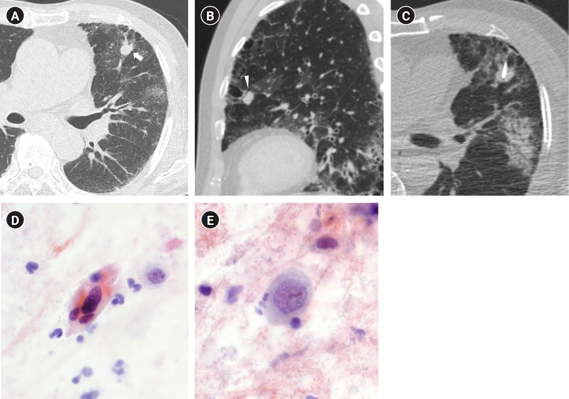
- Using sputum cytology after PCNB to diagnose malignancy, the overall sensitivity, specificity, accuracy, positive predictive value, and negative predictive value were 7.89%, 100%, 20.90%, 100%, and 15.15%. When we compared the diagnostic and non-diagnostic sputum specimen groups, there were no significant differences in age, sex, history of smoking, or pulmonary function testing results. There were significant differences between the groups in lesion size, air-bronchogram in the lesion, multiplicity of lesions, and cell type of squamous cell carcinoma and adenocarcinoma. The lesion size was significantly larger (p=0.001), and the proportion of air-bronchogram (p<0.001) and multiplicity of lesions (p=0.027) was significantly higher in the diagnostic specimen group. The proportion of squamous cell carcinoma (p=0.008) was higher in the diagnostic specimen group, and that of adenocarcinoma was higher in the non-diagnostic specimen group (p=0.012) (Fig. 1). The other lung lesion characteristics (location, solidity, depth from pleura to lesion, presence or absence of necrosis, cavity, mediastinal lymphadenopathy, underlying pulmonary disease of emphysema or fibrosis) and post-PCNB complications such as hemorrhage or pneumothorax did not differ significantly between the groups (Table 3).
- In the multivariate logistic regression analysis, lesion size (odds ratio [OR], 1.035; 95% confidence interval [CI], 1.008–1.064; p=0.013), air-bronchogram in the lesion (OR, 23.485; 95% CI, 2.532–217.316; p=0.005), and cell type of squamous cell carcinoma (OR, 7.397; 95% CI, 1.773–30.865; p=0.006) were significant factors related to the diagnostic sputum specimen after PCNB.
Results
- Cytological or pathological confirmation is essential for diagnosing lung cancer. Several methods for the pathologic evaluation of pulmonary lesions can be used, including CT- or fluoroscopic-guided percutaneous transthoracic lung biopsy, transbronchial biopsy using bronchoscopy with or without endobronchial ultrasound, and surgical resection of lung tissue. However, tissue obtained through a lung biopsy can be non-diagnostic or insufficient, and even in non-diagnostic cases, lung cancer was reported as the final diagnosis in about 40% of cases [17]. Sputum cytology is a simple, noninvasive, and adjuvant method for some patients with large, centrally located tumors. Neumann et al. [18] reported that nearly 50% of all patients with lung cancer who produced adequate sputum specimens were sputum-positive for cancer cells and that the detection of premalignant cells in sputum enabled the early diagnosis of lung cancer.
- The sensitivity and specificity of sputum cytology for detecting lung cancer were 7.89% and 100%, respectively, in this study. Previous studies reported that the overall sensitivity and specificity of sputum cytology were 66% and 99%, respectively, which is a large difference in sensitivity from this study [9]. This difference in sensitivity might reflect the characteristics of the patient groups who underwent PCNB and the process of collecting sputum from those patients. Most patients in this study who underwent PCNB had peripheral lung lesions; few patients with centrally located tumors were included. Also, the sensitivity of sputum cytology in the previous study increased with the number of satisfactory sputum samples tested, starting at 45% for one satisfactory sample, increasing to 55% for two, and rising to 60% for three [19]. In our study, the instruction was to collect only spontaneous sputum without forceful coughing to prevent bleeding after PCNB, which probably affected the sensitivity of the results.
- The lesion size, presence of air-bronchogram, and cell type of lung cancer differed significantly between the non-diagnostic and diagnostic sputum specimen groups.
- A larger lesion size and cell type of squamous cell carcinoma are consistent with the results of previous studies [9,19,20]. Compared with other subtypes of carcinoma, squamous cell carcinoma is mainly located in the airway, grows into the bronchi, and causes atypical changes over a wide area of the respiratory mucosa. Although most lesions in our study were located in the periphery, this characteristic of squamous cell carcinoma might have affected the positive results from sputum specimens.
- Lung cancer with an air-bronchogram was significantly associated with diagnostic sputum in our study. Although the correlation between air-bronchogram and sputum cytology has not been reported in previous studies, we suggest that the presence of a patented bronchus to the lesion affects the sputum cytology results. Atypical cells might emerge into the sputum specimen through the patent open bronchus or airways damaged after PCNB.
- A multiplicity of lung lesions was significantly associated with diagnostic sputum specimens in our study. This result has not been reported previously. By the time the diameter of a lung cancer reaches 1cm, the tumor has more than 109 cells and might have already invaded the bronchial epithelium and vascular epithelium [21]. In cases with multiple lesions or advanced lesions with lung to lung metastases, a multiplicity of lung lesions might be distributed over a larger surface area than a solitary nodule or primary lesion, have high cellularity, and be aggressive.
- Among the commonly known factors, cancer in a central location did not yield significant results in this study. Most of the patients who underwent PCNB in this study had peripheral lung cancers, with few patients with central lung cancer included, which is probably why statistically significant results were not obtained.
- This study had several limitations. First, it was a single-center, retrospective study. Second, only a few patients with diagnostic sputum specimens were included. Among 987 patients, only 177 patients met our inclusion criteria of pathologic confirmation, follow-up, and sputum samples after PCNB for intrapulmonary lesions, and only 12 of those patients produced diagnostic sputum specimens. Despite the challenges in obtaining these specimens, we were trying to analyze factors related to the results in the sputum cytology. The factors not included in the diagnostic sample group, such as part-solid and pure ground glass nodules, might have shown statistical insignificance even if they were actually significant. Third, most of our patients who underwent PCNB were peripheral lung cancer patients. Thus, it is difficult to apply our results to the entire population of lung cancer patients. Fourth, sputum specimens were not obtained through an optimal process. As described above, to prevent bleeding after PCNB, patients were instructed to collect only spontaneous sputum that did not require deep coughing, so the method and number of specimens obtained could not satisfy the optimal environment. In addition, because patients were on bed rest, it is unknown whether the obtained specimens were immediately received in the laboratory. Fifth, we did not conduct a comparison of diagnostic yield between pre- and post- PCNB sputum cytology. Because sputum samples were not routinely obtained before PCNB, it was not possible to compare sputum samples before and after PCNB, so we cannot suggest what effect the PCNB procedure itself had on sputum cytology.
- In conclusion, although post-PCNB sputum cytology has low sensitivity in diagnosing lung cancer, it has shown diagnostic results in some peripheral lung cancer patients who have squamous cell types, relatively large tumors, and air-bronchograms in the lesions. Although this study has many limitations, that is the first study to show the results of post-PCNB sputum analysis and provides insights into the diagnostic yields of post-PCNB sputum cytology in diagnosing lung cancer.
Discussion
-
Conflicts of interest
No potential conflict of interest relevant to this article was reported.
-
Funding
None.
-
Author contributions
Conceptualization: HK. Data curation: SKL. Formal analysis: SKL, HK. Investigation: SKL, HK. Methodology: SKL, HK. Project administration: SKL, HK. Resources: SKL, HK. Software: SKL, HK. Supervision: SP, KNL. Visualization: HK, MJJ. Writing - original draft: SKL. Writing - review & editing: HK, KNL.
Article information

- 1. Ferlay J, Colombet M, Soerjomataram I, Parkin DM, Pineros M, Znaor A, et al. Cancer statistics for the year 2020: an overview. Int J Cancer 2021;49:778–89.ArticlePDF
- 2. Kang MJ, Won YJ, Lee JJ, Jung KW, Kim HJ, Kong HJ, et al. Cancer statistics in Korea: incidence, mortality, survival, and prevalence in 2019. Cancer Res Treat 2022;54:330–44.ArticlePubMedPMCPDF
- 3. Flehinger BJ, Melamed MR, Zaman MB, Heelan RT, Perchick WB, Martini N. Early lung cancer detection: results of the initial (prevalence) radiologic and cytologic screening in the Memorial Sloan-Kettering study. Am Rev Respir Dis 1984;130:555–60.PubMed
- 4. Fontana RS, Sanderson DR, Taylor WF, Woolner LB, Miller WE, Muhm JR, et al. Early lung cancer detection: results of the initial (prevalence) radiologic and cytologic screening in the Mayo Clinic study. Am Rev Respir Dis 1984;130:561–5.PubMed
- 5. Frost JK, Ball WC Jr, Levin ML, Tockman MS, Baker RR, Carter D, et al. Early lung cancer detection: results of the initial (prevalence) radiologic and cytologic screening in the Johns Hopkins study. Am Rev Respir Dis 1984;130:549–54.PubMed
- 6. National Lung Screening Trial Research Team; Aberle DR, Adams AM, Berg CD, Black WC, Clapp JD, et al. Reduced lung-cancer mortality with low-dose computed tomographic screening. N Engl J Med 2011;365:395–409.ArticlePubMedPMC
- 7. Diederich S, Wormanns D. Impact of low-dose CT on lung cancer screening. Lung Cancer 2004;45 Suppl 2:S13–9.ArticlePubMed
- 8. Hubers AJ, Prinsen CF, Sozzi G, Witte BI, Thunnissen E. Molecular sputum analysis for the diagnosis of lung cancer. Br J Cancer 2013;109:530–7.ArticlePubMedPMCPDF
- 9. Schreiber G, McCrory DC. Performance characteristics of different modalities for diagnosis of suspected lung cancer: summary of published evidence. Chest 2003;123(1 Suppl):115S–128S.ArticlePubMed
- 10. Sing A, Freudenberg N, Kortsik C, Wertzel H, Klosa B, Hasse J. Comparison of the sensitivity of sputum and brush cytology in the diagnosis of lung carcinomas. Acta Cytol 1997;41:399–408.ArticlePubMed
- 11. Chae KJ, Hong H, Yoon SH, Hahn S, Jin GY, Park CM, et al. Non-diagnostic results of percutaneous transthoracic needle biopsy: a meta-analysis. Sci Rep 2019;9:12428.ArticlePubMedPMCPDF
- 12. MacMahon H, Naidich DP, Goo JM, Lee KS, Leung AN, Mayo JR, et al. Guidelines for management of incidental pulmonary nodules detected on CT images: from the Fleischner Society 2017. Radiology 2017;284:228–43.ArticlePubMed
- 13. Hansell DM, Bankier AA, MacMahon H, McLoud TC, Muller NL, Remy J. Fleischner Society: glossary of terms for thoracic imaging. Radiology 2008;246:697–722.ArticlePubMed
- 14. Raghu G, Remy-Jardin M, Myers JL, Richeldi L, Ryerson CJ, Lederer DJ, et al. Diagnosis of idiopathic pulmonary fibrosis. An official ATS/ERS/JRS/ALAT clinical practice guideline. Am J Respir Crit Care Med 2018;198:e44–68.ArticlePubMed
- 15. Yun S, Kang H, Park S, Kim BS, Park JG, Jung MJ. Diagnostic accuracy and complications of CT-guided core needle lung biopsy of solid and part-solid lesions. Br J Radiol 2018;91:20170946.ArticlePubMedPMC
- 16. Yamagami T, Yoshimatsu R, Miura H, Yamada K, Takahata A, Matsumoto T, et al. Diagnostic performance of percutaneous lung biopsy using automated biopsy needles under CT-fluoroscopic guidance for ground-glass opacity lesions. Br J Radiol 2013;86:20120447.ArticlePubMedPMC
- 17. Lee KH, Lim KY, Suh YJ, Hur J, Han DH, Kang MJ, et al. Nondiagnostic percutaneous transthoracic needle biopsy of lung lesions: a multicenter study of malignancy risk. Radiology 2019;290:814–23.ArticlePubMed
- 18. Neumann T, Meyer M, Patten FW, Johnson FL, Erozan YS, Frable WJ, et al. Premalignant and malignant cells in sputum from lung cancer patients. Cancer 2009;117:473–81.ArticlePubMed
- 19. Bocking A, Biesterfeld S, Chatelain R, Gien-Gerlach G, Esser E. Diagnosis of bronchial carcinoma on sections of paraffin-embedded sputum. Sensitivity and specificity of an alternative to routine cytology. Acta Cytol 1992;36:37–47.PubMed
- 20. Risse EK, van't Hof MA, Vooijs GP. Relationship between patient characteristics and the sputum cytologic diagnosis of lung cancer. Acta Cytol 1987;31:159–65.PubMed
- 21. Hirsch FR, Franklin WA, Gazdar AF, Bunn PA Jr. Early detection of lung cancer: clinical perspectives of recent advances in biology and radiology. Clin Cancer Res 2001;7:5–22.PubMed
 PubReader
 ePub Link
 Cite
Cite