Outcome of Ductal Carcinoma in Situ in Patients with or Without p53 Mutations
Article information
Abstract
Objectives
p53 is a tumor suppressor gene and plays an important role in the etiology of breast cancer. The aim of this study is to clarify clinical significance of p53 in Ductal Carcinoma in situ (DCIS), and discuss about survival effect.
Methods
The study subjects, 69 women with breast cancer, were a subset of patients operated from Jan 2005 to Dec 2006. We used a cutoff of 10% to distinguish between positive and negative p53 staining. The University of Southern California (USC)/Van Nuys Prognostic Index (VNPI) were compared with 2 categories of p53.
Results
The positivity of p53 was found in 20 patients (29.0%) in DCIS. And negativity of p53 was found in 49 patients (71.0%). And 15 patients (21.7%) had a low USC/VNPI score, 42 patients (60.9%) intermediate and 12 patients (17.4%) a high score. The positivity of p53 was correlated with high USC/VNPI (P = 0.001). The univariate analysis for prognostic factors associated with Disease Free Survival (DFS) revealed that patients with p53 positivity show shorter Disease Free Survival (DFS) than patients with p53 negativity (P = 0.013) and USC/VNPI was also statistically significant (P = 0.030).
Conclusions
According to our study, p53 was associated with high USC/VNPI. These findings suggest that p53 can be used to classify DCIS into at least two subtypes with differing prognoses.
Ductal Carcinoma In Situ (DCIS) of the breast is a complex and heterogeneous spectrum of pathological lesions with a widely variable malignant potential and a not yet clearly understood natural history.1,2 Various molecular markers have been used to predict the prognosis of DCIS.3 Among the molecular marker, the p53 gene is a tumor suppressor gene located in the short arm of chromosome number 17.4 It codes for a nuclear phosphoprotein that appears to play an important role in the regulation of the cell proliferation in that it controls the progression of cells from the G1 to S phase of the cell cycle.5 Mutations in one copy of p53 and/or deletion of the normal gene occur in many human cancers including those of breast, lung, colon, and ovary, astrocytoma, and osteosarcoma, and have been shown to be associated with poor prognosis in breast cancer.6 And the Van Nuys Prognostic Index (VNPI) is a score, developed on the basis of a multivariate retrospective analysis of some measurable prognostic factors in a large series of women.7,8 It is useful in predicting the risk of local recurrence (LR) in patients with conservatively treated DCIS and is therefore an aid to the complex treatment decision-making process.9 This score combines three independent predictors of LR (tumor size, margin width and pathologic classification). The addition of scores (ranging from 1 to 3) from each of these three factors allows the identification of three major groups with low, intermediate and high recurrence risk. The VNPI has been revised as the University of Southern California (USC)/VNPI with the addition of age as a fourth parameter into the scoring system (Table 1). Each group of low (score 4-6), intermediate (score 7-9) or high risk for recurrence (score 10-12) is supposed to be best treated according to these guidelines.10,11 In this paper, we retrospectively applied the new USC/VNPI to our series of DCIS patients treated with surgery, in conjunction with an p53. Particular focus was placed on the role of p53 on disease-free survival.

The USC/VNPI scoring system
MATERIALS AND METHODS
1. Patients and Specimens
Sixty-nine formalin-fixed and paraffin-embedded specimens from patients with pure DCIS were obtained from the Department of OOO at OOO hospital. All of the patients underwent surgery from 2000 to 2005. This retrospective trial was conducted in patients with conservative surgery or mastectomy. Recorded clinical and pathological features for each patient include age, pathologic tumor size, margin status, p53, ER, PgR, HER2 status, surgical treatment and medical adjuvant therapy. Follow-up, including clinical examination (every 6 months for the first 2 years, every one year for the next 3 years) and mammography, bone scan, chest X-ray were carried out in all patients. Recurrence was defined as the first documented evidence of new disease manifestation in the locoregional area, in the contralateral breast, in distant sites, or in a combination of these.
2. Claddification of DCIS
All of the hematoxylin-eosin stained sections of pure DCIS classified by two observers according to the conventional classification methods. The conventional classification was according to the predominant architectural pattern, comedo, cribriform, papillary micropapillary, solid, mixed and so on.12 The Van Nuys Prognostic Index (VNPI) is a score, developed on the basis of a multivariate retrospective analysis of some measurable prognostic factors in a large series of women. It is useful in predicting the risk of local recurrence (LR) in patients with conservatively treated DCIS and is therefore an aid to the complex treatment decision-making process. This score combines three independent predictors of LR (tumor size, margin width and pathologic classification). The addition of scores (ranging from 1 to 3) from each of these three factors allows the identification of three major groups with low, intermediate and high recurrence risk. Several recent papers have shown patient age to be clinically significant. The VNPI has recently been revised as the University of Southern California (USC)/VNPI with the addition of age as a fourth parameter into the scoring system (Table 1). The Van Nuys classification system also defined three groups Each group of low (score 4-6), intermediate (score 7-9) or high risk for recurrence (score 10-12) is supposed to be best treated according to these guidelines.
3. Immunohistochemistry
Immunohistochemical screening for Estrogen receptors (ER), progesterone receptors (PgR) and HER2 status was performed on formalin-fixed paraffin embedded tissue blocks of the primary tumor in the Pathology Department of the OOO. Expressions of p53, ER, PgR and HER2 were determined immunohistochemically on paraffin sections using antibodies against ER, PgR, HER2, p53.13 The pattern of staining was categorised as negative (< 10% of neoplastic cells staining), or positive (> 10% of the neoplastic cells staining).14 The number of p53 protein positive cells in a 4 mm section was expressed as a percentage by counting 500 nuclei of neoplastic cells. ER status and PR status were taken as positive if more than 10% of tumor cells showed staining. Immunohistochemical score of 3+ or FISH + for HER2 was accepted as HER2 positivity.15,16
4.Statistical Analysis
All comparisons between groups and/or parameters were performed using Chi-square test (χ2). A statistical analysis of disease-free survival for each group (obtained by Kaplan-Meier curves for univariate analysis the log rank test) was conducted using the software SPSS 12.0. All P-values were two-sided and a P-value of less than 0.05 was considered to indicate a statistically significant difference. Disease Free survival (DFS) was defined as the time from surgery to first appearance of disease or death for any cause. Survival curves were estimated using the Kaplan-Meier method. Statistical tests were performed using the SPSS 12.0 statistical software package for Windows (SPSS Inc, Chicago, USA).
RESULTS
The main clinicopathological characteristics of the patients in our series are summarized in Table 2. Mean age was 50 years. Tumor size with more 40 mm was reported in 13 patients (18.8%). All patients underwent surgery: conservative surgery was carried out in 45 patients (65.2%) and mastectomy in 24 patients (34.8%). Endocrine therapy was administered to 45 of 69 patients (65.2%). Applying USC/VNPI, 15 patients (21.7%) had a low USC/VNPI, 42 patients (60.9%) intermediate, 12 patients (17.4%) had a high groups. 20 of the 69 patients (29.0%) had positive p53 and 49 patients (71.0%) had negative p53. ER status was positive in 45 patients (65.2%) and negative in 24 patients (34.8%). Older age groups (>60 yrs) were significantly association with Low USC/VNPI (P = 0.010). And the positivity of p53 significantly associated with High USC/VNPI (Table 3) (P = 0.001). No statistical relationship was found between USC/VNPI scores and the other variables, such as PgR, HER2 (Table 4). At a median follow-up of 3.14 years (range, 2.85-3.38). The univariate analysis for prognostic factors associated with DFS revealed that USC/VNPI groups as low, intermediate, high was statistically significant (HR = 2.377: 95%CI of 0.737-7.655, P = 0.030) (Fig. 1) and positivity of p53 was also statistically significant (HR = 2.562: 95%CI of 1.089-6.029, P = 0.0137) (Table 4) (Fig. 2). In our study, USC/VNPI can be used to classify DCIS into at least two subtypes with differing prognosis. And p53 can also be used to predict poor prognosis. As these histological parameters are thought to predict prognosis of DCIS, p53 protein expression may also identify those cases of DCIS which are more likely to progress to invasive carcinoma and therefore influence patient management. Nevertheless the new USC/VNPI is a score easy to calculate and reliable to apply in a clinical setting for predicting the outcome and planning of the therapeutic management of DCIS. We also did not find any statistically significant advantage in groups treated by operation with free wide surgical margins seems to be of better prognostic value than with close margins. But because our study is small size study, more abundant patients' date will be needed to evaluate of the p53's predictive role.
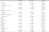
Clinico-pathological features of the patients population and p53 status
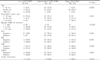
Clinico-pathological features of the patients population and VNPI score distribution

Univariate analysis for variable considered for DFS (Cox proportional hazards regression model)
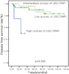
Kaplan-Meier estimates for disease free survival according to USC/VNPI.
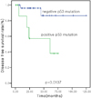
2 Kaplan-Meier estimates for disease free survival according to p53.
DISCUSSION
DCIS is a non-obligate precursor of invasive carcinoma and does not fully express the phenotype of unlimited growth, invasiveness, angiogenesis and metastatic potential.13 The progression to invasive breast cancer (IBC) is likely to result from the accumulation of genetic alterations, allowing clonal selection and the evolution of malignant capability.14 The Van Nuys Prognostic Index (VNPI) is a score, developed on the basis of a multivariate retrospective analysis of some measurable prognostic factors in a large series of women.1 It is useful in predicting the risk of local recurrence (LR) in patients with conservatively treated DCIS and is therefore an aid to the complex treatment decision-making process. This score combines three independent predictors of LR (tumor size, margin width and pathologic classification).7 The addition of scores (ranging from 1 to 3) from each of these three factors allows the identification of three major groups with low, intermediate and high recurrence risk.13,17 Several recent papers have shown patient age to be clinically significant.10 The VNPI has recently been revised as the University of Southern California (USC/VNPI) with the addition of age as a fourth parameter into the scoring system.18 Several papers have analyzed series of patients with DCIS trying to retrospectively evaluate the criteria included by Silverstein in his Van Nuys Prognostic Index as well as other clinical and pathological features, to confirm his value and identify other significant relapse-predictive factors.18,19 The human p53 gene protein is a nuclear phosphoprotein normally expressed at very low levels in all human cells and serves to regulate cell growth and division.20 Direct evidence for the oncogenic capacity of p53 protein has come from the demonstration that it can immortalise and, in co-operation with an activated ras oncogene, transform rodent cells in vitro.13 However, only mutant forms of p53 protein are capable of cell transformation. Furthermore, normally expressed wild-type p53 protein may actually have a tumor suppressor effect. Recent successes in the production of monoclonal antibodies to p53 protein provide valuable tools to detect abnormal expression of the p53 gene product, and high levels of p53 protein expression have been detected in many different types of primary adult carcinomas, including breast, lung, colon, brain, endometrium and bladder.11,21 In breast cancer, it has been shown that p53 protein abnormalities can be present at the in situ phase and that these abnormalities are maintained throughout the stages of breast cancer progression, with no evidence being found for their acquisition during progression of a tumor from in situ to invasive disease or from invasive to metastatic disease. Statistically significant associations were found between p53 protein expression and histological sub-type (P < 0.020), nuclear grade (P < 0.001), and mitotic index (P < 0.050).
Notes
This study was supported by a grant from Kosin University colleage of medicine (2011).