A double-knotted pulmonary artery catheter with large loop in the right internal jugular vein: A case report
Article information
Abstract
Knotting of a pulmonary artery catheter (PAC) is a rare, but well-known complication of pulmonary artery (PA) catheterization. We report a case of a double-knotted PAC with a large loop in a patient with hepatocellular carcinoma (HCC) undergoing liver transplantation, which has been rarely reported in the literature. A PAC was advanced under pressure wave form guidance. PAC insertion was repeatedly attempted and the PAC was inserted 80 cm deep even though PAC should be normally inserted 45 to 55 cm deep. However, since no wave change was observed, we began deflating and pulling the balloon. At the 30-cm mark, the PAC could no longer be pulled. Fluoroscopy confirmed knotting of the PAC after surgery (The loop-formed PAC was shown in right internal jugular vein); thus, it was removed. For safe PA catheterization, deep insertion or repeated attempts should be avoided when the catheter cannot be easily inserted into the pulmonary artery. If possible, the insertion of PACs can be performed more safely by monitoring the movement of the catheter under fluoroscopy or transesophageal echocardiography.
The pulmonary artery catheter (PAC) has been used to monitor hemodynamic changes by measuring cardiac output (CO), pulmonary capillary wedge pressure, and mixed venous oxygen saturation. However, various complications associated with a PAC have been reported. We report a case of double-knotting of a PAC that is one of the complications of pulmonary artery (PA) catheterization.
CASE
A 40-year-old man (weight, 64 kg; height, 174.2 cm) was admitted to our institution for liver transplantation for hepatocellular carcinoma (HCC). This patient had undergone liver resection twice before that led to recurrences. He had no other underlying disease, and his echocardiogram and laboratory results were within normal range.
Anesthesia was induced via an intravenous injection of 14 mg etomidate and 20 mg cisatracurium, and intubation was performed. For monitoring continuous arterial blood pressure, cannulation was performed on the right radial artery and the dorsalis pedis artery. The femoral vein and the right internal jugular vein (IJV) were cannulated under ultrasound guidance. Particularly, an advanced venous access high-flow device (AVA HF, Edwards Lifesciences LLC, Irvine, California, USA) was used for the cannulation of the right IJV, through which a PAC (Swan-Ganz CCOmbo V, Edwards Lifesciences LLC, Irvine, California, USA) was inserted.
Although the entry of the PAC into the right ventricle (RV) was confirmed through pressure monitoring via the distal port of the PAC, it could not be inserted into the PA. After 3 attempts with a maximal depth of 50 cm, surgery was started while monitoring CO with FloTracTM. We did not tried any other attempts, including using fluoroscopy or transesophageal echocardiography (TEE) and changing position because of the inexperience of the practitioner. During surgery, we attempted to insert the PAC. After 2 attempts, the PAC was inserted 80 cm deep, incidentally. However, since no wave and electrocardiogram changes were observed, we started deflating and pulled the balloon. At the 30-cm mark, the PAC could no longer be pulled. We decided to examine the PAC with fluoroscopy after surgery and remove it.
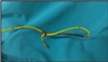
The fluoroscopic image revealed a double-knotted catheter with a large loop in the right IJV (Fig. 1). A small skin incision was made at the site of PAC insertion, and a venotomy with a purse-string suture was performed on the right IJV by surgeon. Then, the PAC was removed by pulling it through an opening in the center (Fig. 2). The patient was discharged without any complications.

Fluoroscopy after the surgery shows a knotted pulmonary artery catheter with loop formation.
Knotted pulmonary artery catheter removed by venotomy.
DISCUSSION
The PAC known as the Swan-Ganz catheter, developed by Swan, Ganz, and their colleagues in the 1970s, is used to measure cardiac output, pulmonary capillary wedge pressure, and mixed venous oxygen saturation. It allows for the observation and control of various hemodynamic changes and is still used.1 However, its use has been controversial because it can cause complications such as a balloon rupture, pulmonary infarction, PA rupture, arrhythmias, RV perforation, and knot formation.2 Although there are non-invasive devices for measuring cardiac output, such as lithium dilution cardiac output (LiDCO™), pulse contour cardiac output (PiCCO®), and FloTrac™, their accuracy to measure CO is inferior to that of the PAC with significant aortic regurgitation, arrhythmia, and they have produced different clinical treatment outcomes.3 Therefore, the PAC has been consistently used despite its risks.
At our institution, a PAC is generally used for monitoring cardiac output and other hemodynamic parameters.
Knotting of a PAC occurs at a rate of 0.2% to 2.5%, commonly from inserting the PAC 60 cm or deeper without a PA wave or repeatedly performing insertion and withdrawal. Furthermore, a PAC accounts for two-thirds of all intravascular knotted catheters due to its thin and soft wall and long shape.4 To prevent knots, it is important to carefully monitor insertion under pressure wave form guidance. If no PA wave is observed after insertion by an additional 20 cm after observing an RV wave, or if no PA wave appears even after the PAC is inserted 60 cm deep, the balloon must be removed, and the PAC must be gradually pulled back. Changing the patient's position may more effectively prevent PAC knotting.5 With advances in imaging technology, a PAC insertion can be performed aided by other imaging methods. A PAC insertion can be performed under fluoroscopy guidance, which produces real-time animated images using an x-ray, or TEE, which allows for continuous viewing of the cardiac chamber using ultrasound. These methods may enable safer and more accurate positioning of the PAC.56 However, fluoroscopy- or TEE-guided PAC insertion has been not performed at our institution routinely.
Once a knotted intravascular catheter occurs, various methods can be used to remove it. A PAC can be pulled while a sheath is fixed to reduce the knot size, and then the sheath and PAC can be removed via the insertion site.7 An intervention can be used in which a snaring catheter or wire is used to remove the knot.8 Surgical removal can also be performed.9 Removal of a knotted catheter via an intervention is the safest method because it is less invasive than surgical removal, reduces the knot size, and is less likely to cause vascular trauma; therefore, it has been increasingly used. Herein, the catheter was double knotted with a large loop, and a tail that formed as the tip of the catheter curved, posing a high risk for vascular damage. Because the knot was right beneath the skin, surgical removal was deemed more efficient than an intervention, and was consequently performed. The catheter was removed without any difficulty.
In summary, to prevent knot formation, a catheter must be sufficiently inserted without balloon inflation, and the use of a catheter with a diameter that is too small should be avoided. Insertion of PACs can be performed more safely by monitoring the movement of the catheter under fluoroscopy or TEE. Deep insertion or repeated attempts should be avoided when the catheter cannot be inserted into the PA. Physicians should be more careful about performing PAC insertions in patients who have RV enlargement, and are more prone to knotting10 or risk factors such as old age, female sex, pulmonary hypertension, coagulation disorders, anticoagulation therapy, and hypothermia.11