Transient Inferior Oblique Muscle Palsy Following Transconjunctival Lower Lid Blepharoplasty
Article information
Abstract
Although the inferior oblique (IO) muscle is positioned considerably deep in the orbit, transconjunctival lower lid blepharoplasty may affect it and transient or permanent IO muscle palsy might result. Therefore diplopia should be explained before cosmetic blepharoplasty performed with transconjunctival approach.
With advanced age, the supportive force of the orbital septum of the palpebra becomes weak, and the fat located beneath the orbital septum escapes and protrudes in an irregular manner. When the symptoms are severe, the patient may be negatively impacted aesthetically, psychologically, and socially; thus, lower lid blepharoplasty may be performed. Compared with transcutaneous lower lid blepharoplasty, transconjunctival lower lid blepharoplasty is aesthetically superior since no scars are left on the skin and less lid retraction occurs. Moreover, this procedure better exposes the operational view and the duration of the operation is short. Therefore, transconjunctival lower lid blepharoplasty has been more frequently performed in recent times, particularly among those patients whose fat alone has protruded.1
Inferior oblique muscle palsy following transconjunctival lower lid blepharoplasty has been reported in rare cases internationally, but no case report of such an occurrence has previously been published in South Korea.1234 Herein, the authors report a case of monocular inferior oblique muscle palsy, which occurred following transconjunctival lower lid blepharoplasty, and that spontaneously improved 2 months later.
CASE
A 34-year-old male patient who had undergone binocular transconjunctival lower lid blepharoplasty in a private plastic surgery clinic 3 weeks earlier presented to our institution with the chief complaint of diplopia, which occurred immediately following blepharoplasty. The patient complained that diplopia occurred particularly when he gazed in both the upper and right upper direction, but he had no diplopia in other directions. The patient was a healthy person and he did not have a medical history of strabismus, ocular injury, ocular surgery, or any other systemic diseases. The naked vision was 1.0 in the right eye and 1.0 in the left eye. The manifest refraction test results were −0.5 DSph in the right eye and emmetropia in the left eye. The patient's intraocular pressure was 13 mmHg in the right eye and 15 mmHg in the left eye. The near vision was 1.0 in both eyes. The Titmus stereoscopic vision test result was normal. Pupilloscopy showed a normal direct light reflex result. The pupil size was the same in both eyes. The slit-lamp biomicroscopy showed normal results in the anterior segment of the eye, including the conjunctiva and the conjunctival sac. The visual field test showed orthophoria with respect to far distances and near distances, but the elevation of the left eye was limited to −1. The alternate prism cover test results were 14 prism diopters (PD) hypotropia in the left eye and 5 PD esotropia in the right upper direction, 5 PD hypotropia in the left eye and 5 PD esotropia in the upper direction, and 5 PD esotropia in the left upper direction (Fig. 1). The remaining results were 3 PD hypotropia in the left eye during right gaze, 4 PD hypotropia in the left eye upon head tilt, and orthophoria in the other directions, including during left head tilt. The Lancaster test showed type A esotropia accompanying binocular incyclotropia and left-eye hypotropia (Fig. 2). The double Maddox rod test showed five-degree incyclotropia in the left eye. In addition, funduscopy showed a finding that was indicative of incyclotropia in both eyes (Fig. 3). The Worth four-dot test showed a normal result at the original position of eyes, but diplopia was evident in both the upper and right upper direction. The forced duction test result was normal.
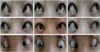
Nine gaze extraocular movement photography show left hypotropia in right upper and upgaze at the initial presentation (white arrows).

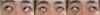
Lancaster test demonstrates esotropia and intorsion in both eyes, and left hypotropia at the initial presentation.

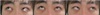
Fundus photography show suspicious intorsion in both eyes at the initial visit.
Orbital computed tomography (CT), which was performed to differentiate between the mechanical damage caused by lower lid blepharoplasty from myasthenia gravis and dysthyroid ophthalmopathy, showed no anatomical abnormalities in the extraocular muscle or the orbital wall. The acetylcholine receptor antibody assay, the repetitive nerve stimulation test (Jolly test), and the thyroid function test also showed normal results. Therefore, the patient was diagnosed with inferior oblique muscle palsy of the left eye.
The alternate prism cover test performed 1 week later showed improved results of 10 PD hypotropia in the left eye and 3 PD esotropia in the right upper direction, 4 PD hypotropia in the left eye and 3 PD esotropia in the upper direction, and 3 PD hypotropia in the left eye during right head tilt, as well as orthophoria in all other directions (Fig. 4).
Extraocular movement photography demonstrate limitation of elevation at right upper gaze in the left eye 1 month following blepharoplasty (white arrow).
The alternate prism cover test performed 2 months following the operation showed orthophoria in all directions, except for 2 PD hypotropia in the left eye in the right upper direction (Fig. 5). The double Maddox rod test showed no cyclodiplopia. The Lancaster test showed improvements in the esotropia, hypotropia, and incycloduction. The diplopia disappeared in all gaze directions.
Extraocular movment photography show decreased limitation of elevation at right upper gaze in the left eye 2 month following blepharoplasty (white arrow).
DISCUSSION
Lower lid blepharoplasty has recently, and extensively, been performed as an aesthetic surgery. Transconjunctival lower lid blepharoplasty does not leave a scar on the skin and causes fewer adverse effects, such as lower lid retraction, ectropion, and illacrimation, when compared with transcutaneous lower lid blepharoplasty. However, transconjunctival lower lid blepharoplasty may result in direct damage to the inferior oblique muscle or the inferior rectus muscle, causing diplopia or strabismus.12345
Syniuta et al.3 analyzed 12 cases of patients whose diplopia continued for 3 months or longer due to acquired strabismus following transconjunctival lower lid blepharoplasty; the authors reported that inferior rectus muscle palsy was found in seven patients, and partial amputation of the inferior oblique muscle was found in two patients, one of whom also exhibited partial amputation of the inferior rectus muscle. Ghabrial et al.1 analyzed six patients; four showed injury at both the inferior oblique muscle and the inferior rectus muscle, and one showed an injury at the lateral rectus muscle. Moreover, the researchers reported that two of the patients required strabismus correction surgery, while the remaining four patients recovered during the follow-up, suggesting that the cause of strabismus in these patients may have been intramuscular hemorrhage and edema, the generation of intramuscular scar tissue, and/or the insertion of the extraocular muscle between the tarsal membranes.
Considering the mechanism that causes inferior oblique muscle palsy during transconjunctival lower lid blepharoplasty from an anatomical perspective, it seems that the inferior oblique muscle starts to run from the lacrimal fossa, while the first one-third of the inferior oblique muscle runs to the lateral posterior part and passes through the capsulopalpebral fascia and the lower part of the inferior rectus muscle to form the middle third of the muscle. Finally, the lateral third of the inferior oblique muscle wraps around the eyeball to adhere to the lateral sclera. The orbital fat of the lower lid is divided by the inferior oblique muscle into the inner fat and the central fat; the central fat and the lateral fat are separated by the arcuate expansion of the inferior oblique muscle membrane.6 Even when the first third of the inferior oblique muscle is damaged, permanent damage to the inferior oblique muscle function is rare because the middle third, where the capsulopalpebral fascia and Lockwood's ligament crosses, serves as a secondary point of origin. Therefore, during transcutaneous lower lid blepharoplasty, even when the first third of the inferior oblique muscle is damaged while the inner fat is removed (particularly because the tissue near the nose is not well exposed during the skin incision), inferior oblique muscle palsy rarely develops due to the existence of the secondary point of origin. Conversely, for transconjunctival lower lid blepharoplasty, the incision starts from the posterior conjunctival sac, and thus the middle third of the inferior oblique muscle may be damaged if fat is removed without figuring out the position of the inferior oblique muscle, resulting in permanent palsy of the inferior oblique muscle.2 As a method to prevent the development of inferior oblique muscle palsy, Syniuta et al.3 recommended performing the conjunctival incision at a location 3 to 4 mm of the lower boundary of the tarsal plate, while Frankel2 suggested that the incision be made 5 mm of the lower boundary of the tarsal plate; after the dissection of the anterior part should be performed, dissection toward the lower part of the inferior tarsal muscle be carried out. Also, Ghabrial et al.1 stated that the conjunctival incision performed at the region 10 mm lower to the corneal limbal is safe, and that the rotation of the eyeball should be verified by carefully moving the eyeball with the forceps when removing the fat tissue. Klapper et al.7 stated that the temporary post-operational extraocular muscle malfunction may be caused by traumatic intramuscular hemorrhage and edema, or muscular toxicity due to the use of a local anesthetic; therefore, the Hess test or the Lancaster test should be performed if there is diplopia. If the diplopia does not disappear after 8 weeks of follow-up, prism eyeglasses should be prescribed; similarly, re-operation - including a strabismus repair operation - should be considered if the diplopia does not improve after a few months.
The patient in the present case improved within 2 months; this was likely due to the fact that, first, the inferior oblique muscle palsy may have resulted from slight intramuscular hemorrhage and edema, since the position and size of the inferior oblique muscle, the adjacent connective tissues, and the orbital wall were found to be normal during orbital CT with a uniform intramuscular shadow. The intramuscular injection of the local anesthetic, which was limited to the inferior oblique muscle, may have intensified the inferior oblique muscle palsy. Second, the inferior oblique muscle palsy may have been temporary since the inferior oblique muscle remained normal, except for the first third of the inferior oblique muscle, which was partially damaged during the conjunctival incision followed by dissection. Third, the local anesthetic might have caused partial muscular toxicity, which later disappeared.
Direct damage to the inferior oblique muscle rarely occurs when a surgeon operates deep inside the conjunctiva and removes a considerable amount of fat tissue without verifying the inferior oblique muscle; this is related to excessive electrocautery and dissection, as well as to muscular hemorrhage.4 In the case of direct damage to the inferior oblique muscle, a strabismus repair surgery may be required; however, the patient may continue to suffer strabismus even after the surgery due to either the synechia of the scar tissues or to the muscular trauma.
It is necessary to carefully observe the state of the orbital wall and the orbital muscle by performing orbital radiography in order to differentiate between the inferior oblique muscle palsy and other diseases that cause inferior oblique muscle malfunction. Since there has been a case report of strabismus caused by extraocular muscle pulleys of the inferior rectus muscle due to scar tissues, it can be concluded that if there is an accompanying vertical strabismus, then the anatomical position and thickness of the muscle, as well as the synechia and the positional movement of the extraocular muscle pulley, should be taken into consideration.8
In conclusion, since permanent strabismus resulting from transconjunctival lower lid blepharoplasty is very rare, yet still possible, a surgeon should perform the operation accurately, while understanding the anatomical structures of the eyelid, the extraocular muscle, and the orbit. The surgeon should also provide the patient with sufficient explanations regarding the possibility of diplopia and strabismus development prior to the operation. In addition, an ophthalmologist should consider the possibility of extraocular muscle damage in a patient with diplopia following transconjunctival lower lid blepharoplasty. Herein, the authors reported a case of inferior oblique muscle palsy following transconjunctival lower lid blepharoplasty, which has not yet been reported domestically, and also provided a review of the relevant literature.