Expression of vascular endothelial growth factor is a clinically useful predictor for aggressive basal cell carcinoma
Article information
Abstract
Objectives
Basal cell carcinoma (BCC) tumors are locally invasive but rarely metastatic. However, aggressive metastatic variants are being increasingly reported in elderly people. Here we investigated the clinical utility of vascular endothelial growth factor (VEGF) as a predictive biomarker for aggressive BCC variants.
Methods
Thirty-five pathologically confirmed cases of BCC that underwent surgical removal in the Plastic Surgery Department between January 1, 2011 and December 31, 2012 were studied. VEGF expression was analyzed in formalin-fixed paraffin-embedded tumor tissue by immunohistochemical staining. Positive staining was defined as more than 10% of the tumor cells showing immunoreactivity. The associations of VEGF expression with various clinicopathologic parameters were analyzed.
Results
The face was the most prevalent site (28/35), with 15 cases from the nose, 6 cases from the eyelid, and 5 cases from the cheek. The patients were aged between 41 and 86 years, with a mean age of 69.26 ± 173.903 years. The mean BCC size was 1.34 ± 3.853 cm, with a range of 0.3 cm to 12.0 cm. The mean tumor invasion depth from the basement epidermal membrane was 0.17 ± 0.035 cm, with a range of 0.03 cm to 1.10 cm. A mean of 5.66 ± 20.938 intraoperative frozen section slides were examined. VEGF was not expressed in 14 of the 35 patients (40.0%), whereas 42.9% of the patients had low expression and 17.1% of the patients had high expression. VEGF expression was significantly associated with age (P = 0.022), size (P = 0.030), site (P = 0.013), tumor invasion depth (P = 0.019), and number of intraoperatively frozen sections (P = 0.003).
Conclusions
These results suggest that VEGF expression as assessed by immunohistochemistry can predict aggressive or poor prognosis in BCC.
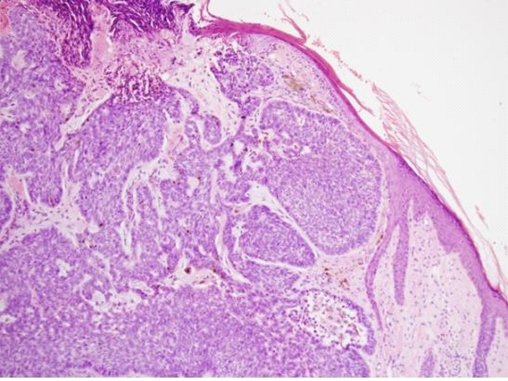
The pathologic findings of Basal cell carcinoma of skin on the face. (H-E, x100)
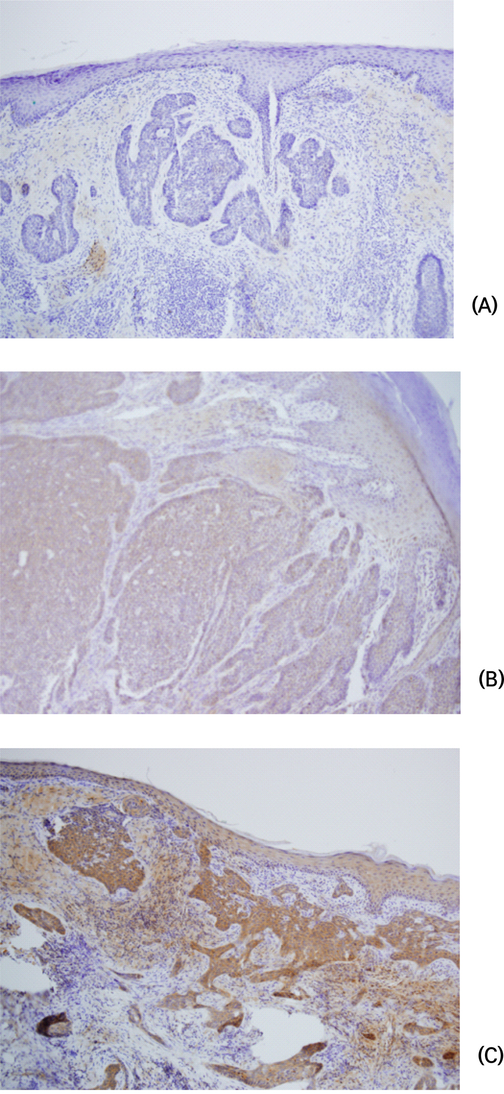
The expression of VEGF by immunostaining in the basal cell carcinoma (x100) (A) Negative expression of VEGF in the basal cell carcinoma (B) Low-expression of VEGF in the basal cell carcinoma (C) High-expression of VEGF in the basal cell carcinoma

The association of VEGF expression with clinicopathologic variables in the basal cell carinoma