Giant Right Atrial Thrombus associated with Tunneled Cuffed Hemodialysis Catheter: A Case of Successful Treatment with Thrombolytic Agent and Anticoagulant

Article information

Kosin Med J. 2013;28(1):61-65
Publication date (electronic) : 2013 January 19
doi : https://doi.org/10.7180/kmj.2013.28.1.61
1Department of Internal Medicine, College of Medicine, Hallym University, Seoul, Korea
2Hallym Kidney Research Institute, College of Medicine, Hallym University, Seoul, Korea
Corresponding author: Young-Ki Lee, Department of Internal Medicine, Hallym Kidney Research Institute, Hallym University College of Medicine, 948-1, Daelim-dong, Yeongdeungpo-gu, Seoul 150-950, Korea TEL: +82-2-829-5214 FAX: +82-2-848-9821 E-mail: km2071@unitel.co.kr
Received 2012 December 23; 2012 December 26; Accepted 2013 January 07.

Abstract

There are a variety of tunneled cuffed hemodialysis catheter-related complications including infection, thrombus formation, and catheter dysfunction. Catheter-related thrombus in right atrium is a rare complication and treatment guideline for atrial thrombus does not exist. A 3.0×2.8 cm sized giant atrial thrombus was found in a 35-year-old female hemodialysis patient. She was treated with catheter removal, thrombolysis and anticoagulation therapy. Size of atrial thrombus was gradually decreased and left ventricular systolic function was clearly improved after treatment. We experienced and reported a case of giant right atrial thrombus associated with tunneled cuffed hemodialysis catheter that was successful treated with thrombolytic agent and anticoagulant.
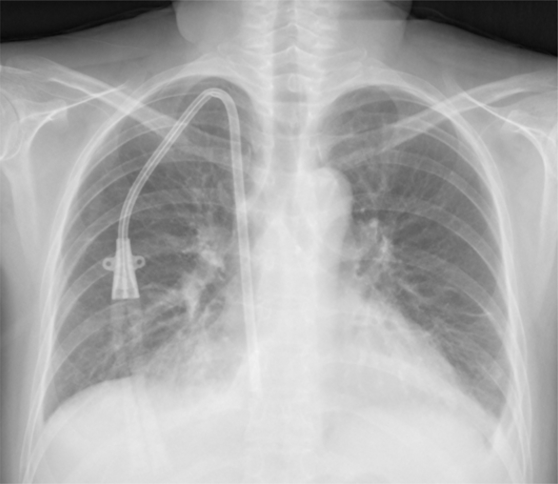
Fig. 1.

Chest PA shows cardiomegaly, bilateral pleural effusion and a central venous catheter. Tip of catheter through right internal jugular vein is located at right atrium.
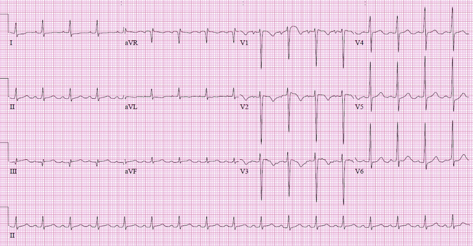
Fig. 2.

Electrocardiography shows T-wave inversion in lead V1-3. But there in no significant change for previous study.
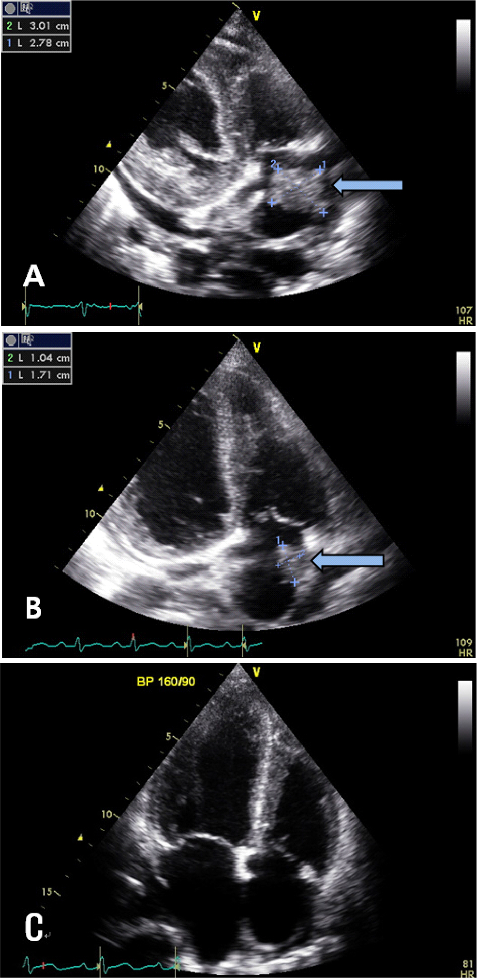
Fig. 3.

(A) Transthorasic echocardiography shows a 3.0×2.8 cm sized mass in right atrium on admission. (B) After 2 months, echocardiography shows a 1.0×1.7 cm sized mass. (C) After 6 months, echocardiography shows no mass in right atrium.
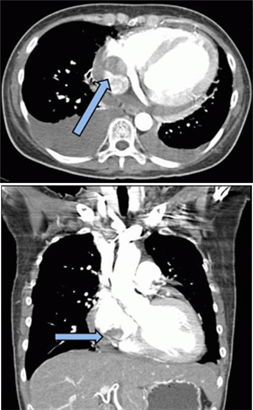
Fig. 4.

Chest CT agiography shows low-attenuated filling defect in the right atrium (arrow), attached to the tunneled cuffed hemodialysis catheter.

References

1. Vascular Access 2006 Work Group. Clinical practice guidelines for vascular access. Am J Kidney Dis 2006;48:S176–247.
2. Ghani MK, Boccalandro F, Denktas AE, Barasch E. Right atrial thrombus formation associated with central venous catheters utilization in hemodialysis patients. Intensive Care Med 2003;29:1829–32.
3. Fuchs S, Pollak A, Gilon D. Central venous catheter mechanical irritation of the right atrial free Wall: A cause for thrombus formation. Cardiology 1999;91:169–72.
4. Lee BH, Kim HG, Lee JY, Kang BK, Chung ES, Seo KI, et al. Huge infected thrombus of right atrium related to hemodialysis catheter. Korean J Nephrology 2009;28:514–8.
5. Ram R, Swarnalatha G, Rakesh Y, Jyostna M, Prasad N, Dakshinamurty KV. Right atrial thrombus due to internal jugular vein catheter. Hemodial Int 2009;13:261–5.
6. van Laecke S, Dhondt A, de Sutter J, Vanholder R. Right atrial thrombus in an asymptomatic hemodialysis patient with mal-functioning catheter and patent foramen ovale. Hemodial Int 2005;9:236–40.
7. Kinney EL, Wright RJ. Efficacy of treatment of patients with echocardiographically detected right-sided heart thrombi: a meta-analysis. Am Heart J 1989;118:569–73.
8. Negulescu O, Coco M, Croll J, Mokrzycki MH. Large atrial thrombus formation associated with tunneled cuffed hemodialysis catheters. Clin Nephrol 2003;59:40–6.
9. Rose PS, Punjabi NM, Pearse DB. Treatment of right heart thromboemboli. Chest 2002;121:806–14.
10. Horner SM, Bell JA, Swanton RH. Infected right atrial thrombus--an important but rare complication of central venous lines. Eur Heart J 1993;14:138–40.

Article information Continued

Fig. 1.

Chest PA shows cardiomegaly, bilateral pleural effusion and a central venous catheter. Tip of catheter through right internal jugular vein is located at right atrium.

Fig. 2.

Electrocardiography shows T-wave inversion in lead V1-3. But there in no significant change for previous study.

Fig. 3.

(A) Transthorasic echocardiography shows a 3.0×2.8 cm sized mass in right atrium on admission. (B) After 2 months, echocardiography shows a 1.0×1.7 cm sized mass. (C) After 6 months, echocardiography shows no mass in right atrium.

Fig. 4.

Chest CT agiography shows low-attenuated filling defect in the right atrium (arrow), attached to the tunneled cuffed hemodialysis catheter.