A Case of Endobronchial Actinomycosis with a Broncholith cured by Cryotherapy through a Flexible Bronchoscope
Article information
Abstract
We report the case of a 53-year-old man who presented with obstructive pneumonitis and broncholithiasis. We attempted to remove the broncholith with forceps through a flexible endoscope, but the potential for bleeding due to partial synechia did not allow this. We succeeded in removing it with cryotherapy. The histopathological diagnosis was thoracic actinomycosis associated with broncholithiasis. Endobronchial actinomycosis with a broncholith is very rare. We successfully treated a patient with endobronchial actinomycosis with a broncholith by administering short-term antibiotics after broncholithectomy via cryotherapy through a flexible bronchoscope.

Chest radiograph shows focal infiltration in the right middle lobe.
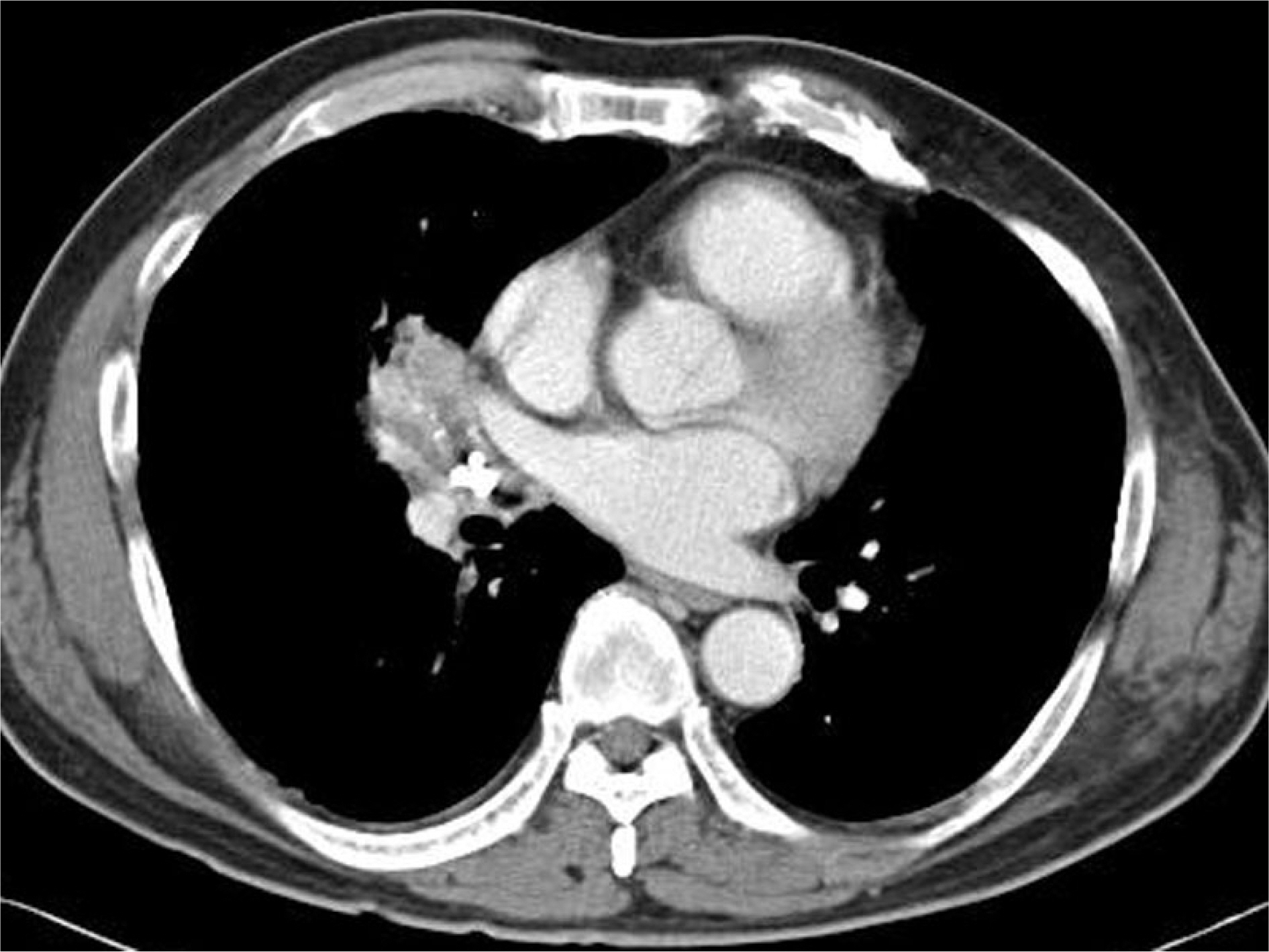
Chest CT shows a calcified stone in the bronchus and atelectasis in the right middle lobe.

Bronchoscopy shows near-total obstruction of the right middle lobe by broncholith.

There were no specific findings at follow-up bronchoscopy 5 days after the cryotherapy procedure.

The bronchoscopic biopsy showed marked lymphocyte infiltration and a few calcified fragments with radiating filamentous basophilic bacterial colonies (H&E, × 40).

There were no specific findings at follow-up bronchoscopy 8 weeks after the cryotherapy procedure.