KOSIN UNIVERSITY COLLEGE OF MEDICINE
Articles
- Page Path
- HOME > Kosin Med J > Volume 36(2); 2021 > Article
-
Case Reports
Hemangioma in Renal Pelvis - Myeong Su Chu
-
Kosin Medical Journal 2021;36(2):175-179.
DOI: https://doi.org/10.7180/kmj.2021.36.2.175
Published online: December 31, 2021
Department of Internal Medicine, Jeil Yeonhap clinic, Sejong-si, Republic of Korea
- Corresponding Author: Myeong Su Chu, Department of Internal Medicine, Jeil Yeonhap clinic, 62-15, Saeromjungang-ro, Sejong-si, 30127, Republic of Korea, Tel: +82-44-715-5501, Fax: +52-44-715-5503, E-mail: chooms80@hanmail.net
• Received: October 18, 2021 • Revised: October 18, 2021 • Accepted: November 1, 2021
Copyright © 2021 by Korean Association of Medical Journal Editors
Articles published in Kosin Medical Journal are open-access, distributed under the terms of the Creative Commons Attribution Non-Commercial License (http://creativecommons.org/licenses/by-nc/4.0/) which permits unrestricted non-commercial use, distribution, and reproduction in any medium, provided the original work is properly cited.
- 1,220 Views
- 11 Download
- 1 Crossref
Abstract
- Hemangioma in the renal pelvis is a very rare benign tumor that may be mistaken for renal cell carcinoma. We present, herein, a case of a 59-year-old woman with a renal mass that was diagnosed as a cavernous hemangioma in the renal pelvis. The patient underwent intravenous pyelography, urine cytology, retrograde pyelography, kidney dynamic computed tomography (CT), and surgical excision.
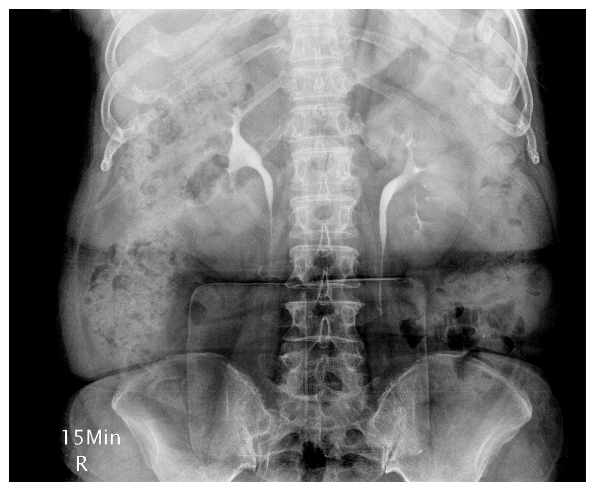
- A 59-year-old female patient with a painless gross hematuria that started 5 days before being admitted to a private hospital was taken from the hospital for a computed tomography . The patient was then transferred to this hospital for precise examination and treatment. According to her medical history, she had been taking medication for hypertension from another hospital for 10 years and there was nothing unusual in her family history. Based on the examination findings, she was alert with a height of 155 cm, weight of 65 Kg, blood pressure of 130/80 mmHg and pulse rate of 76/min. Her breathing rate was 20/min and her temperature was 37.0 C. There were no abnormal findings in heart sound and respiratory sound, abdominal auscultation, palpation and side abdominal percussion. In the laboratory tests, there were no unusual findings in a complete blood test and blood chemistry tests. In the urinalysis many/high-power fields (HPF) and WBC 0–1/HPF were observed. From the intravenous pyelography (IVP) performed, a filling defect in the middle calyx of the left kidney was suspected (Fig. 1), and blood leakage in the left ureteral opening was observed from the cystoscope. The result of a urine cytology test was negative. A retrograde pyelography (RGP) confirmed the filling defect in the middle calyx of the left kidney (Fig. 2). Kidney dynamic computed tomography (CT) was carried out and a poorly enhanced 3.5 × 4.5 cm diameter round local mass was observed in the middle calyx of the left kidney (Fig. 3). Therefore, the authors performed a left nephrectomy as indicated for the treatment of kidney cancer. The post-op progress was good, and it was diagnosed as hemangioma in the left renal pelvis from the pathological biopsy (Fig. 4).
CASE
- A hemangioma is a vasculogenic tumor which can be divided into benign hemangioendothelioma, capillary hemangioma, cavernous hemangioma, venous hemangioma, and racemose hemangioma.1 A cavernous hemangioma is mostly found in the skin or the surface of a mucous membrane, and occasionally it is found in the liver, spleen, pancreas or kidney.1,2 It usually affects people between the ages of 30 to 40 years old. Both the ratio between men and women and the left and right ratio are the same. Although most cavernous hemangioma occur unilaterally, 12% occur in multiple.3 In general, cavernous hemangioma occurs alone in the urinary tract, but rarely it can be accompanied by multiple sclerosis, Sturge-Weber syndrome, or Klippel Trenaunay-Weber syndrome.3,4 The size is usually reported as about 1 to 2 cm and it may have a similar diameter with the kidney size.3,4
- Clinical symptoms can include intermittent or persistent hematuria, and about 40% of patients complain of pain when passing the hematoma through the urinary tract.4 Hemangioma of the kidney, although it is benign, may cause serious bleeding and may obstruct the ureteropelvic border. 4 Clinically, the key is that benign masses like hematoma in the kidney is to be able to distinguish them from malignant masses.
- During urinary angiography, if the size of the mass is big, defects in contrast medium shading of the renal calyx or renal pelvis, or deformation of the renal calyx, may occur due to protrusion or pressure; but if the size is smaller, the technique may show normal findings.4 Abdominal ultrasonography of cavernous hemangioma typically shows a border with a low echo shadow, and unlike renal cell carcinoma, it does not have an acoustic shadow. However, it is often difficult to distinguish cavernous hemangioma from renal cell carcinoma with ultrasonography. Abdominal computed tomography of cavernous hemangioma shows enhanced contrast, but it is a non-specific finding.4
- Partial nephrectomy is the principal course of treatment for cavernous hemangioma. However, since it is difficult to distinguish cavernous hemangioma in the kidney from a malignant renal tumor in practice, it is often diagnosed only after radical nephrectomy.5 If the diagnosis is confirmed before surgery, endoscopic treatment can be performed and in the case of healthy people who show mild hematuria but do not show any other clinical problems, observing the progress is not contraindicated.5
- All cases of cavernous hemangioma in the kidney reported in Korea were diagnosed after nephrectomy. In this case presented by the authors, nephrectomy was performed since it was difficult to distinguish it from renal cell carcinoma, and as a result, it could be diagnosed as hemangioma of kidney.
- The characteristics of this authors’ case is that among the cases reported in Korea, the size was found to be the largest at 3.5 to 4.5 cm, unlike the previously reported cases of 1 to 2 cm.
DISCUSSION

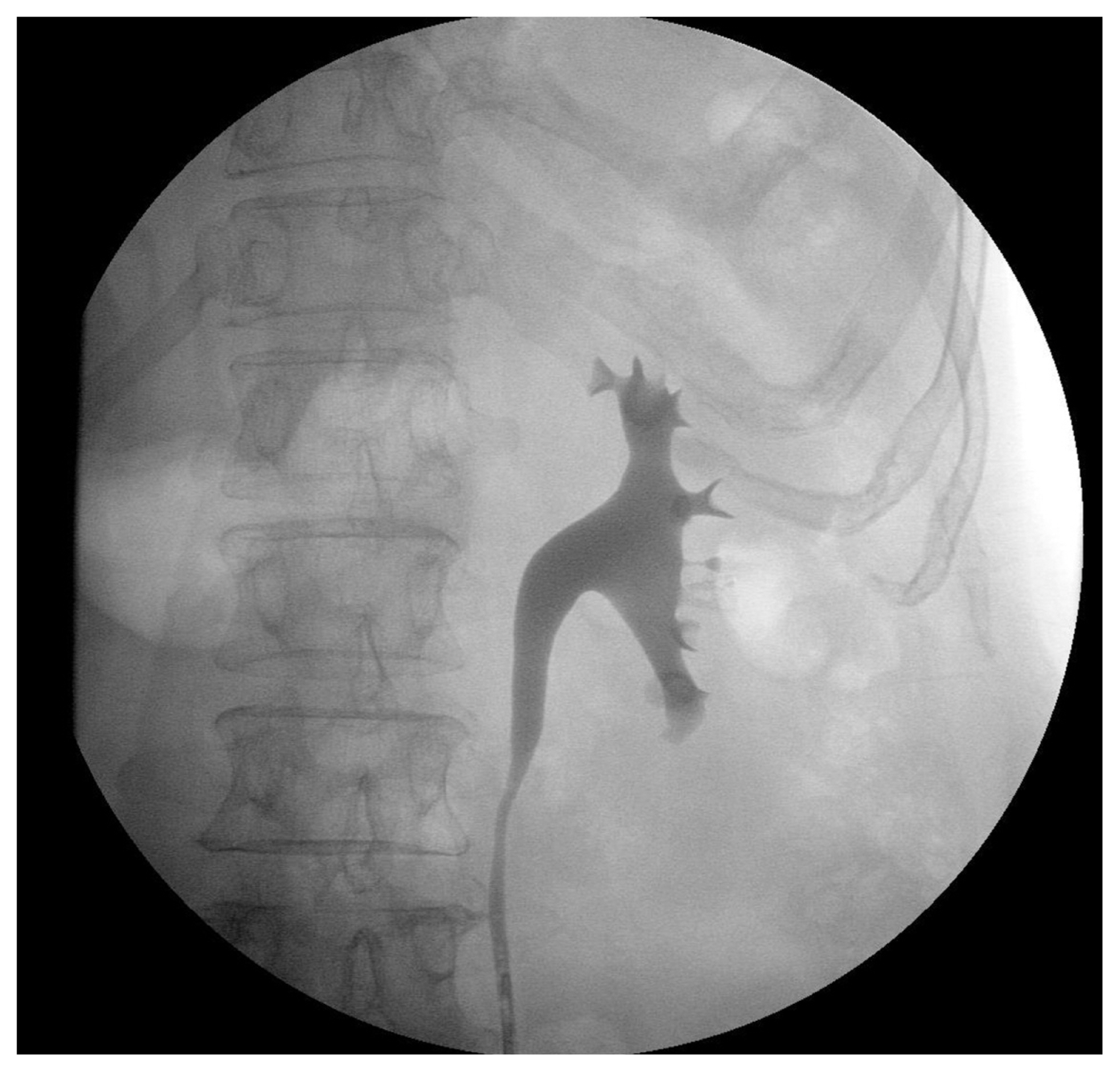
Fig. 3(A) Axial kidney dynamic computed tomography (CT) images show a poorly enhanced mass-like lesion in the upper calyx of the left kidney (arrow). (B) Coronal kidney dynamic CT images show a poorly enhanced masslike lesion in the upper calyx of the left kidney (arrow).
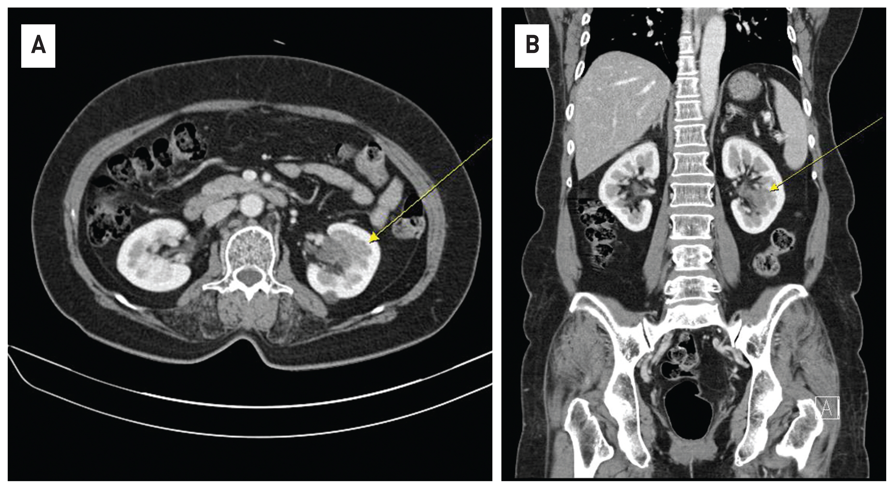
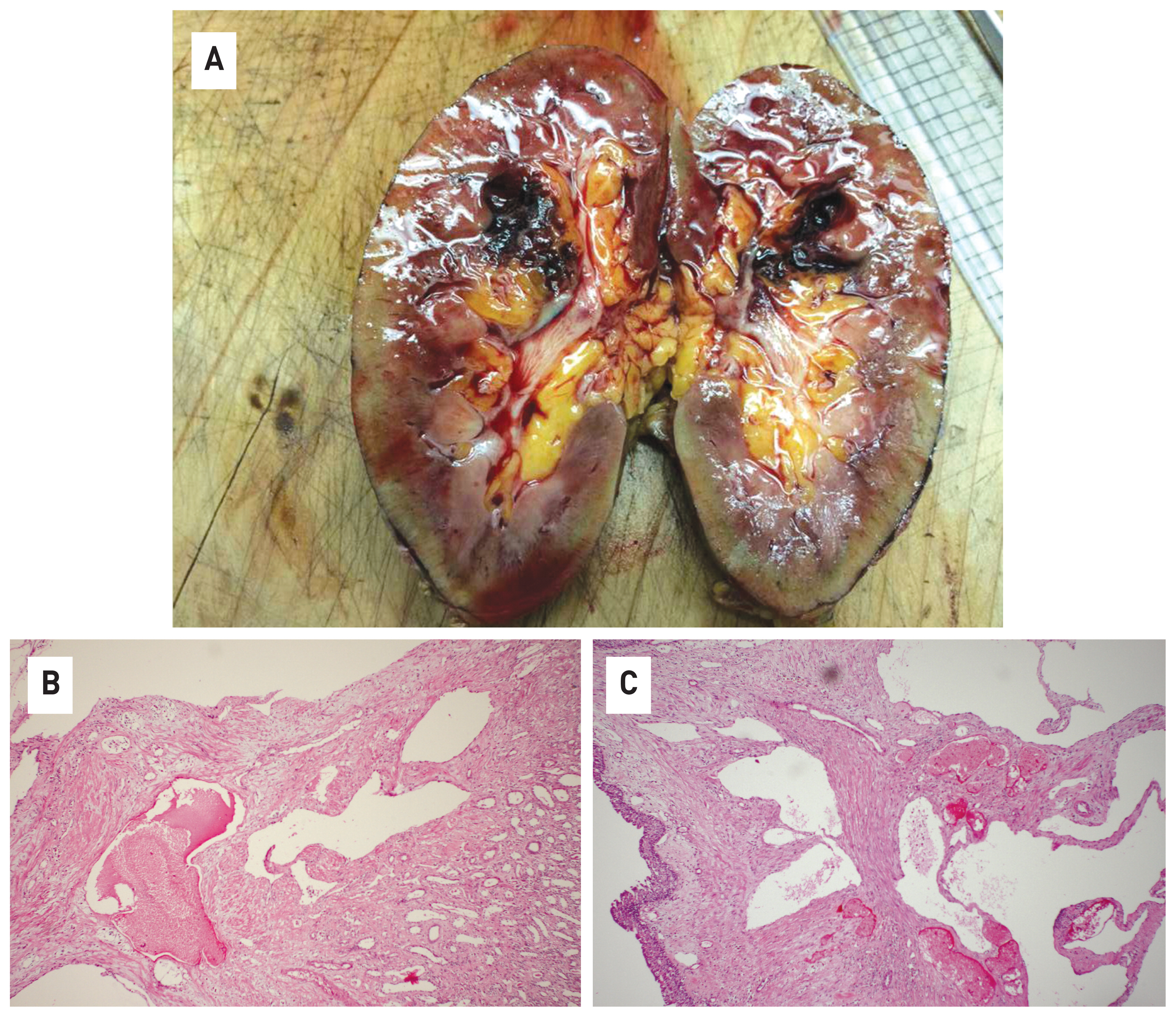
Fig. 4(A) The macroscopic finding shows a 3.5x4.5 cm hemorrhagic, sponge-like round mass in the pole of the left kidney. (B) The microscopic finding shows dilated blood-filled vessels (H&E satin, x100). (C) The microscopic finding shows dilated blood-filled vessels (H&E satin, x100).

- 1. Jahn H, Nissen HM. Haemangioma of the urinary tract: review of the literature. Br J Urol 1991;68:113–7.ArticlePubMed
- 2. Haas CA, Resnick MI, Abdul-Karim FW. Cavernous hemangioma presenting as a renal hilar mass. J Urol 1998;160:2139–40.ArticlePubMed
- 3. Peterson NE, Thompson HT. Renal hemangioma. J Urol 1971;105:27–31.ArticlePubMed
- 4. Lee HS, Koh BH, Kim JW, Kim YS, Rhim HC, Cho OK, et al. Radiologic findings of renal hemangioma: report of three cases. Korean J Radiol 2000;1:60–3.ArticlePubMedPMC
- 5. Daneshmand S, Huffman JL. Endoscopic management of renal hemangioma. J Urol 2002;167:488–9.ArticlePubMed
References
Figure & Data
References
Citations
Citations to this article as recorded by 
- Renal venous malformation misdiagnosed as carcinoma: A report of one case and the review of literature
Xinchun Zhang, Ning Wang, Bingkui Yang
International Journal of Immunopathology and Pharmacology.2023;[Epub] CrossRef
 PubReader
 ePub Link
 Cite
Cite