KOSIN UNIVERSITY COLLEGE OF MEDICINE
Articles
- Page Path
- HOME > Kosin Med J > Volume 36(1); 2021 > Article
-
Case Report
Right Atrial Blood Cyst Mimicking a Vegetative Mass - Sun Hack Lee, Jung Hyun Choi
-
Kosin Medical Journal 2021;36(1):40-43.
DOI: https://doi.org/10.7180/kmj.2021.36.1.40
Published online: June 30, 2021
Department of Cardiology, Pusan National University Hospital, Pusan National University School of Medicine, Busan, Korea
- Corresponding Author: Jung Hyun Choi, Department of Cardiology, Pusan National University Hospital, 179, Gudeok-ro, Seo-gu, Busan 49241, Korea, Tel: +82-51-240-7514 Fax: +82-51-240-7763 E-mail: mariahyeon@gmail.com
• Received: April 17, 2020 • Revised: July 20, 2020 • Accepted: January 17, 2021
Copyright © 2021 by Korean Association of Medical Journal Editors
Articles published in Kosin Medical Journal are open-access, distributed under the terms of the Creative Commons Attribution Non-Commercial License (http://creativecommons.org/licenses/by-nc/4.0/) which permits unrestricted non-commercial use, distribution, and reproduction in any medium, provided the original work is properly cited.
- 932 Views
- 9 Download
Abstract
- A 79-year-old woman presented to another hospital with complaints of right leg pain. Computed tomography and magnetic resonance imaging of the spine was performed in the other hospital, which showed an abscess in the right iliacus muscle. She was referred to our hospital because of a mass in the right atrium on echocardiography. Inflammatory markers were elevated, and Staphylococcus aureus were identified in blood cultures. Transthoracic echocardiography revealed a shaggy mass in the right atrium that resembled vegetation. Transesophageal echocardiography showed a large cystic mass with a hyperechoic lesion. After surgery, biopsy results indicated that it was a myxoid mass with cystic changes.
- A 79-year-old woman presented to another hospital with complaints of right leg pain for one week. Abdominal computed tomography (CT) and spinal magnetic resonance imaging (MRI) was performed at that hospital, which showed an abscess in the right iliacus muscle. She was referred to our hospital because of a mass in the RA detected by echocardiography. At the time of admission, an electrocardiogram showed normal sinus rhythm with a right bundle branch block, and all cardiac markers were within normal range. Inflammatory markers were elevated (white blood cell, 22,750 /μL, C-reactive protein 19.14 mg/dL), and Staphylococcus aureus was identified in blood cultures.
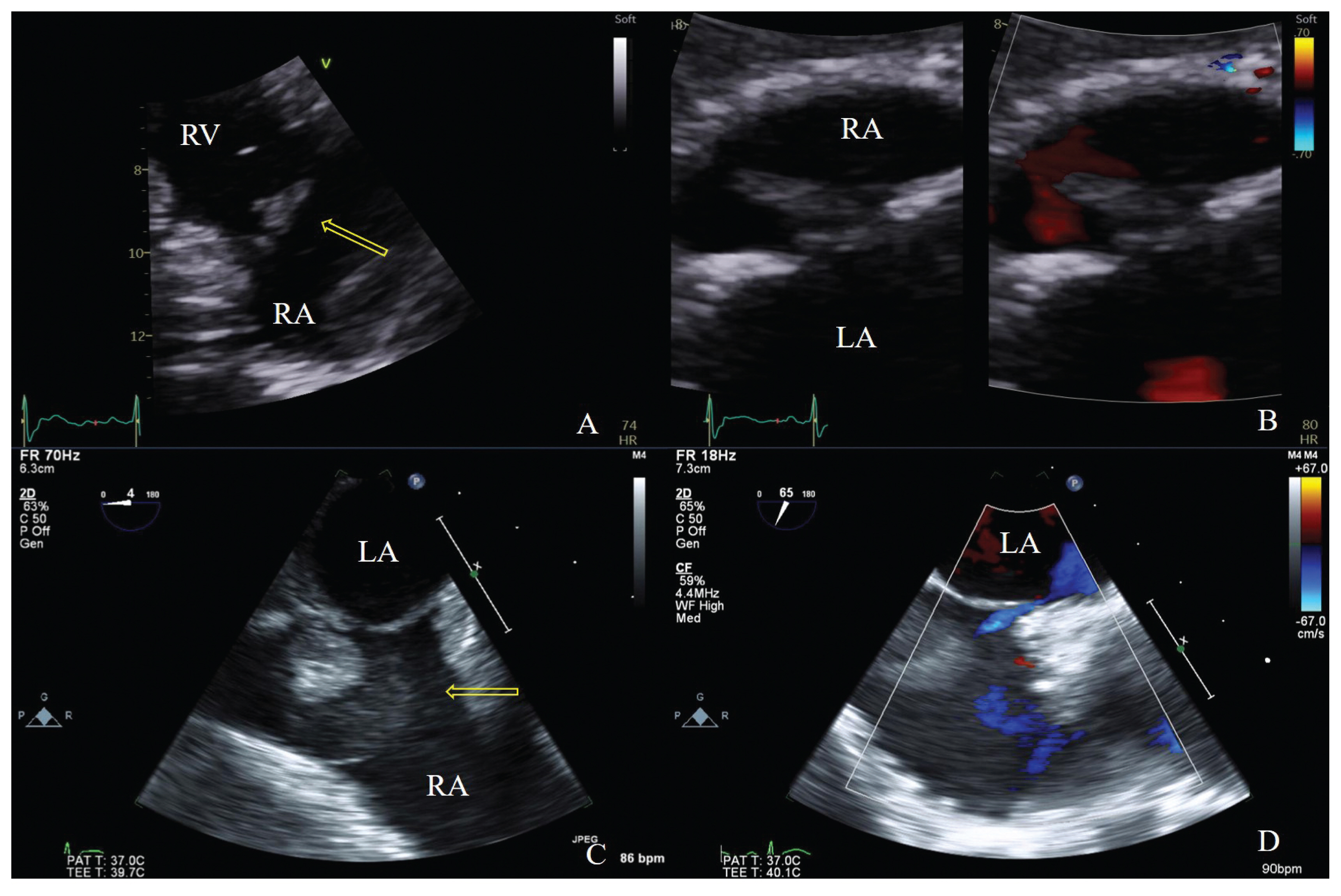
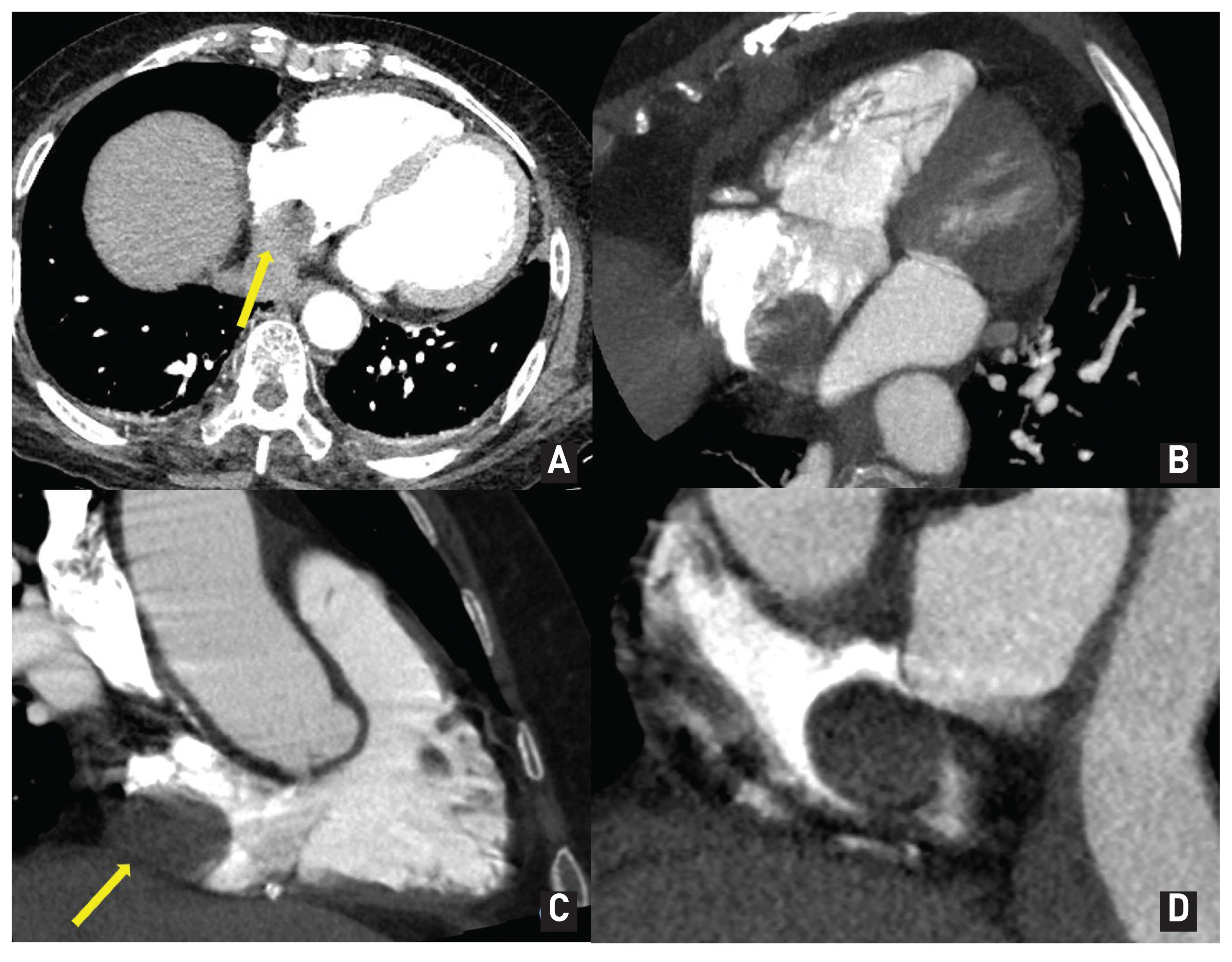
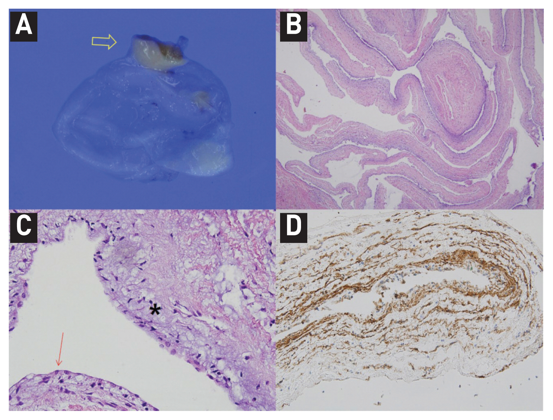
- Transthoracic echocardiography (TTE) at our hospital revealed a hypermobile mass (1.7 × 0.6 cm) on the interatrial septum in the RA, similar to the previous examination which was performed at another hospital. We thought that it was a vegetation (yellow arrow, Fig. 1A, 1B). However, transesophageal echocardiography (TEE) showed another aspect to the mass, a hypoechoic mass. It was 1.9 × 1.7 cm and looked like a myxoma or cyst (yellow arrow, Fig. 1C). The hyperechoic mass, which appeared on TTE, was attached to the surface of that mass. A patent foramen ovale was confirmed by color Doppler (Fig. 1D). The shunt flowed toward the RA mass, where there was hyperechoic mass. We thought that it was a newly developed vegetation on the previously existing RA mass. A cardiac CT image appeared to show a myxoma, which was the RA mass without enhancement (Fig. 2). The differential diagnosis of this mass included a blood cyst, myxoma, and vegetation. We surgically removed the mass, which was pathologically confirmed as a myxoid with cystic changes (Fig. 3). There was no evidence of vegetation on the surface of the cyst. The inflammatory marker was improved gradually as the abscess in the right iliacus muscle was improved.
CASE
- Primary benign cardiac tumors are rare, and approximately 50% are myxomas. Other benign cardiac tumors, including cysts, have an incidence of 0.0017% in the general population. Intracardiac cysts are observed less frequently than pericardial cysts.2,5 Blood cysts in the heart were first reported in 1844 by Elsasser. 6 These cysts are congenital in origin and rare in adults. Cardiac blood cysts are typically found on the closure of the valvular endocardium, but can grow in all heart cavities.3,7 A patient with a blood cyst, high fever, and chills was previously reported by Dumantepe et al.8 In that report, the patient’s fever was attributed to a pulmonary embolism and there was no bacteremia.
- We encountered a difficult case where a blood cyst mimicked vegetation in a patient with Staphylococcus aureus bacteremia. There are no recommendations for the treatment of asymptomatic blood cysts. Some authors have monitored the cyst size and found that it did not increase in size over time.9 However, in this case, the patient had fever and bacteria were identified in repeated blood cultures; therefore, surgical treatment was necessary.
DISCUSSION
Fig. 1
(A) TTE in the parasternal RV inflow view showing a shaggy-appearing mass. (B) TTE in the subxiphoid view, showing a mass with color Doppler. (C) TEE shows a large hypoechoic mass and shaggy-appearing mass. (D) A patent foramen ovale is seen on TEE.
TTE, transthoracic echocardiography; RV, right ventricular; TEE, transthoracic echocardiography

Fig. 2
Heart CT image showing a 2.5 cm mass arising from the interatrial septum of the RA.
CT, computed tomography; RA, right atrium

Fig. 3
(A) Gross findings of the resected RA mass. The cystic mass shows a translucent cystic wall without a solid nodule. The pedicle of the cystic mass is identified (arrow). (B) Microscopic examination of the resected specimen. A. At low magnification, the cystic wall shows myxoid changes in the surface aspect and is supported by a fibromuscular wall (H-E stained, x40). (C) At a high-power view (x400), the cystic wall appears composed of the flat surface epithelium and subepithelial myxoid stroma (flat surface epithelium; red arrow, myxoid stroma; star). (D) The surface epithelium and stellate or spindle-shaped stromal cells appear focally positive for CD34 which was often positive in myxoma10.
RA, right atrium; H-E, hematoxylin and eosin

- 1. Roberts WC. Primary and secondary neoplasms of the heart. Am J Cardiol 1997;80:671–82.ArticlePubMed
- 2. Peters PJ, Reinhardt S. The echocardiographic evaluation of intracardiac masses: a review. J Am Soc Echocardiogr 2006;19:230–40.ArticlePubMed
- 3. Shakerian B, Jebelli M. Right Atrium Blood Cyst and Calcified Kernel in an Adult. Clin Med Insights Case Rep 2019;12:1–2.Article
- 4. Park MH, Jung SY, Youn HJ, Jin JY, Lee JH, Jung HO. Blood cyst of subvalvular apparatus of the mitral valve in an adult. J Cardiovasc Ultrasound 2012;20:146–9.ArticlePubMedPMC
- 5. Lee WC, Huang MP, Fu M. Multiple intracardiac masses: myxoma, thrombus or metastasis: a case report. J Med Case Rep 2015;9:179.ArticlePubMedPMC
- 6. Elsässer C. Bericht uber die ereignisse in der gebaranstalt des CatherinenHospital in Jahre 1844. Med Correspondenzblatt 1844;14:297.
- 7. Otsuka H, Arinaga K, Fukuda T, Takaseya T, Shojima T, Takagi K, et al. Double Right Atrial Blood Cysts. Ann Thorac Surg 2016;101:e147–9.ArticlePubMed
- 8. Dumantepe M, Ak K, Mungan U, Alp I, Inan BK, Yilmaz AT. Blood cyst of the right ventricle presenting as recurrent high fever and chills in an adult. Ann Thorac Surg 2009;87:638–40.ArticlePubMed
- 9. Agac MT, Acar Z, Turan T, Karadag B, Kul S, Erkan H. Blood cyst of tricuspid valve: an incidental finding in a patient with ventricular septal defect. Eur J Echocardiogr 2009;10(5):88–9.Article
- 10. Wang JG, Li YJ, Liu H, Li NN, Zhao J, Xing XM. Clinicopathologic analysis of cardiac myxomas: Seven years’ experience with 61 patients. J Thorac Dis 2012;4:272–83.PubMedPMC
References
Figure & Data
References
Citations
Citations to this article as recorded by 
 PubReader
 ePub Link
 Cite
Cite